Module 1.2
Anterior Transverse: Subscapularis
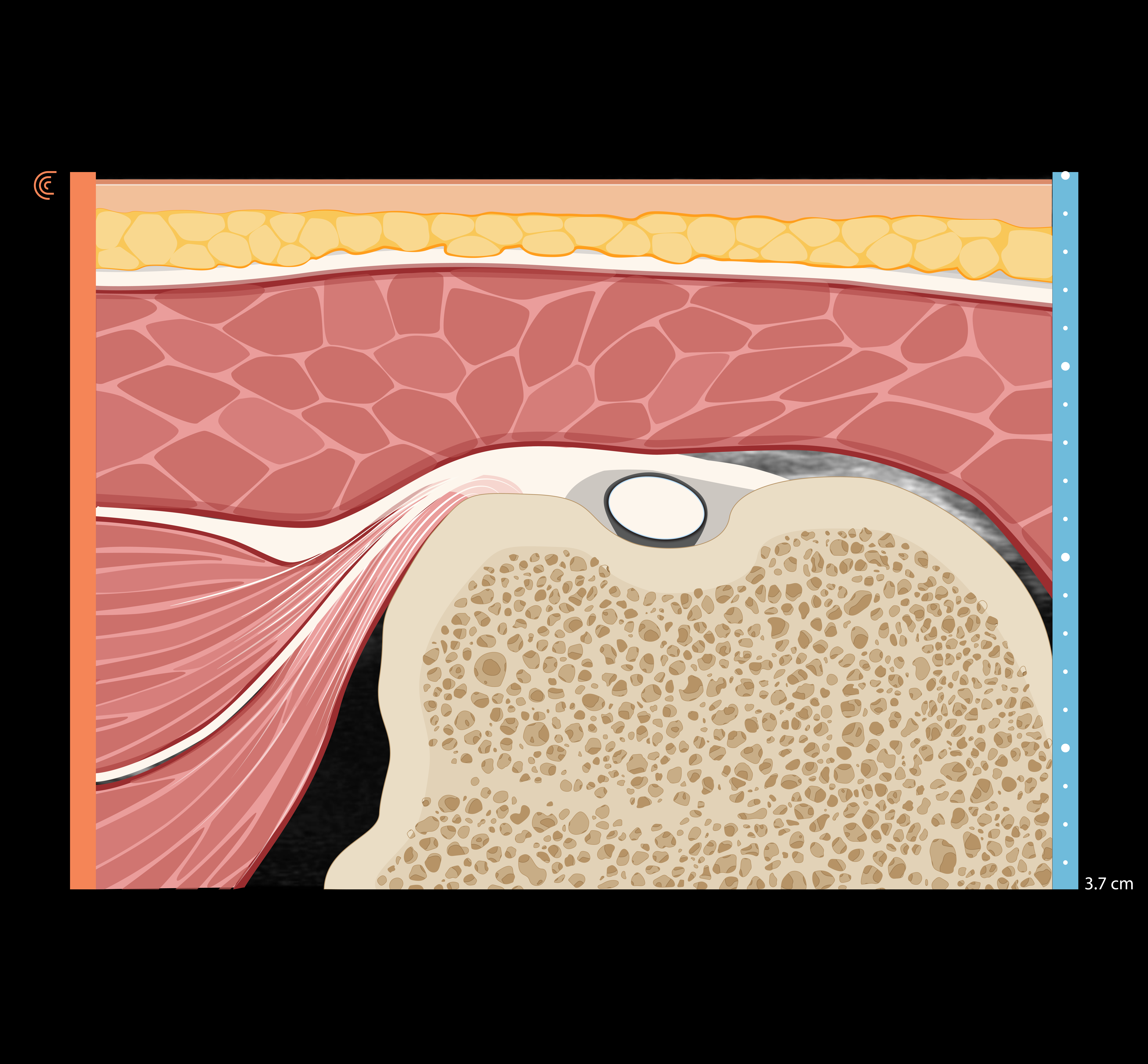
In this section, we will use ultrasound during internal and external shoulder rotation to explore subscapularis and its insertion on the lesser tubercle. Structures visualized will include the greater and lesser tubercles of the humerus, the deltoid, the long head of the biceps brachii, the subscapularis tendon, and the coracoid process.
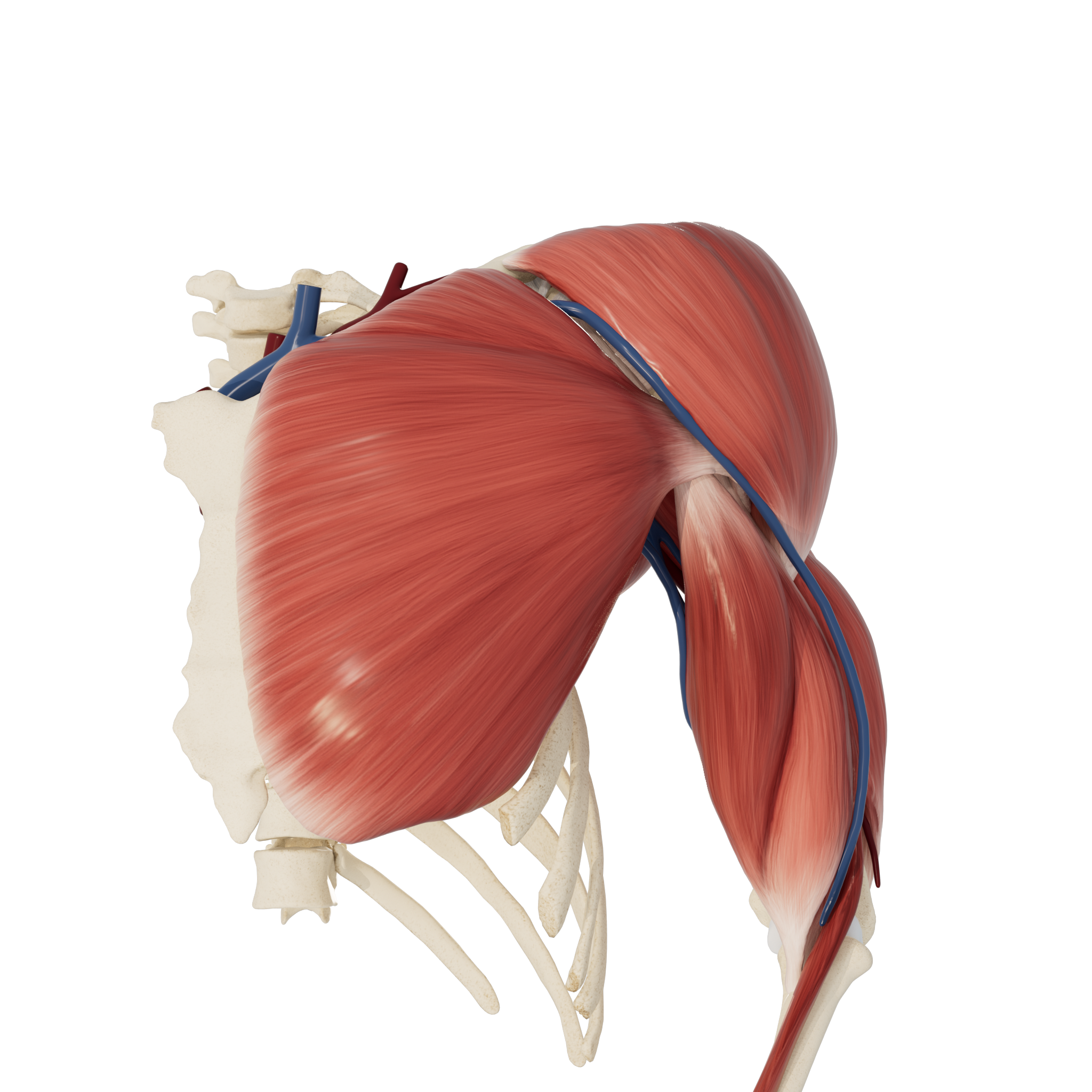
Patient starting position:
The patient’s arm should be relaxed and in neutral position.
Note: this examination is demonstrated on the left shoulder, with the ultrasound probe indicator facing medially to begin. The numbers used to denote each step on the model correspond to the direction of the indicator. The orientation marker is always on the left side of the sonogram.
Patient starting position
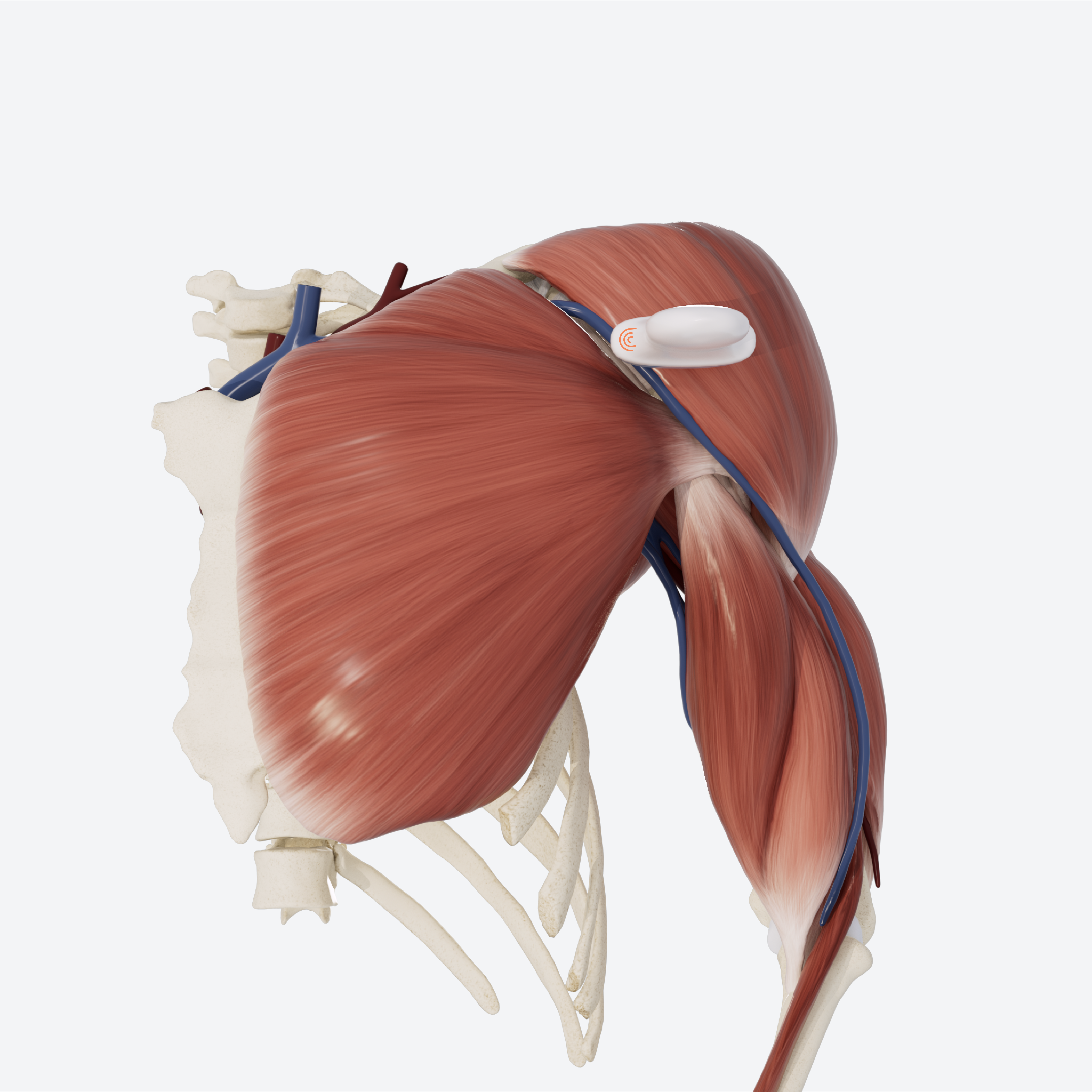
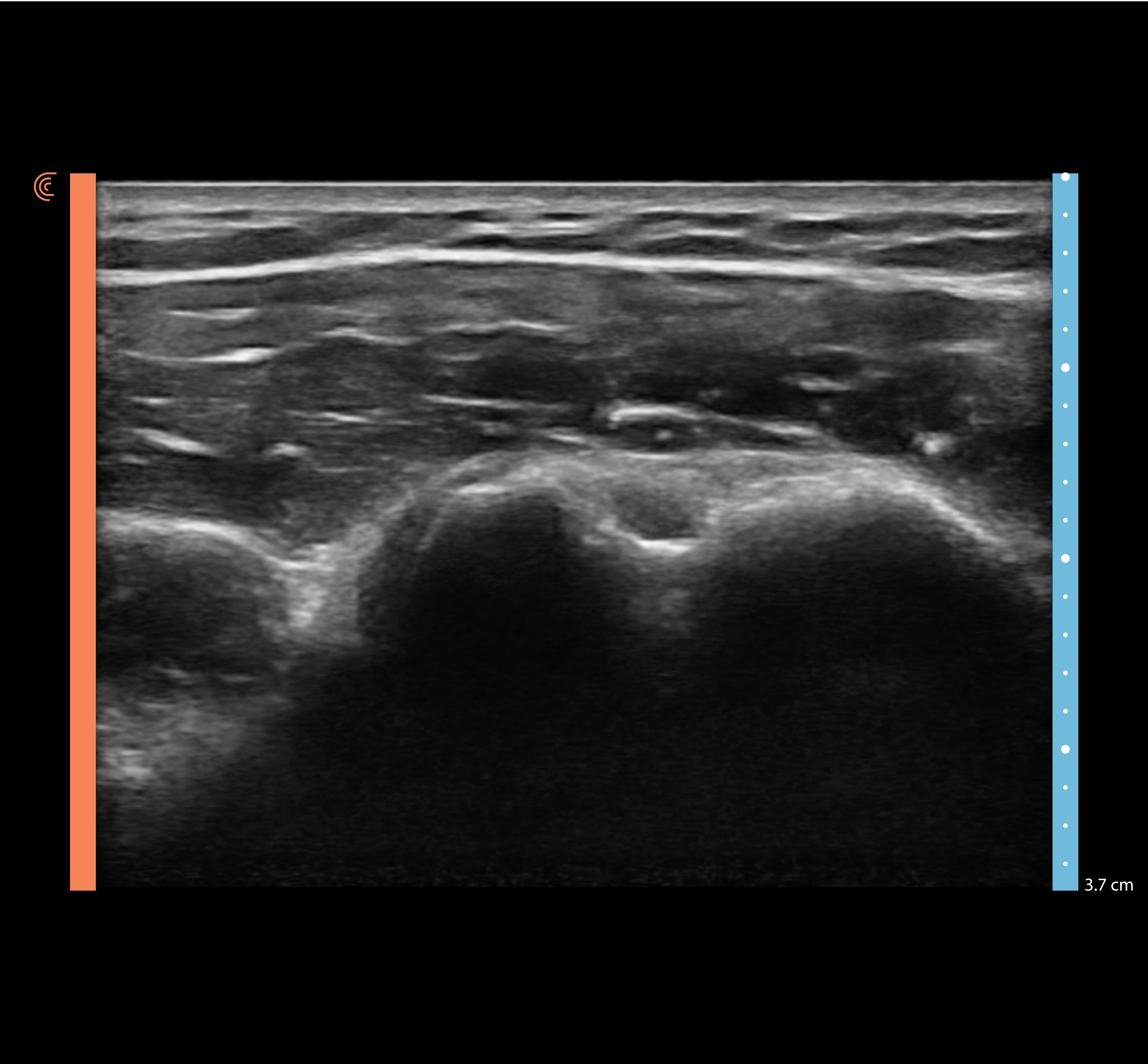
STEP 1 Bicipital Groove in Internal Rotation
Begin with bicipital groove visualized in transverse orientation. You should be able to locate the greater and lesser tubercles.
STEP 2 Subscapularis: Long Axis
While keeping the probe still, have the patient externally rotate their arm with their forearm slightly supinated and their elbow anchored against the torso. This will stretch subscapularis into view. Slide the probe superiorly and inferiorly on the tendon to view insertion width on the lesser tubercle.
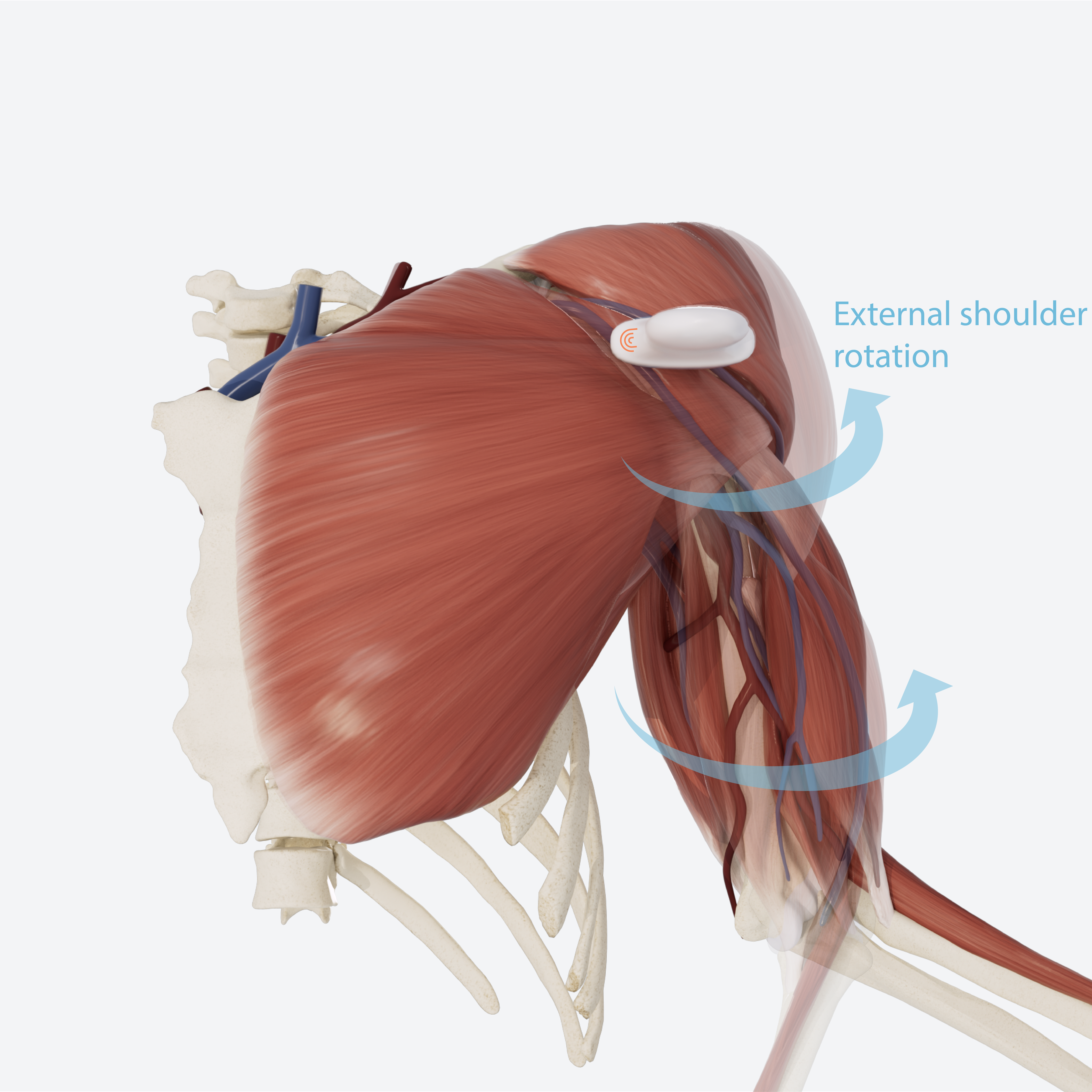
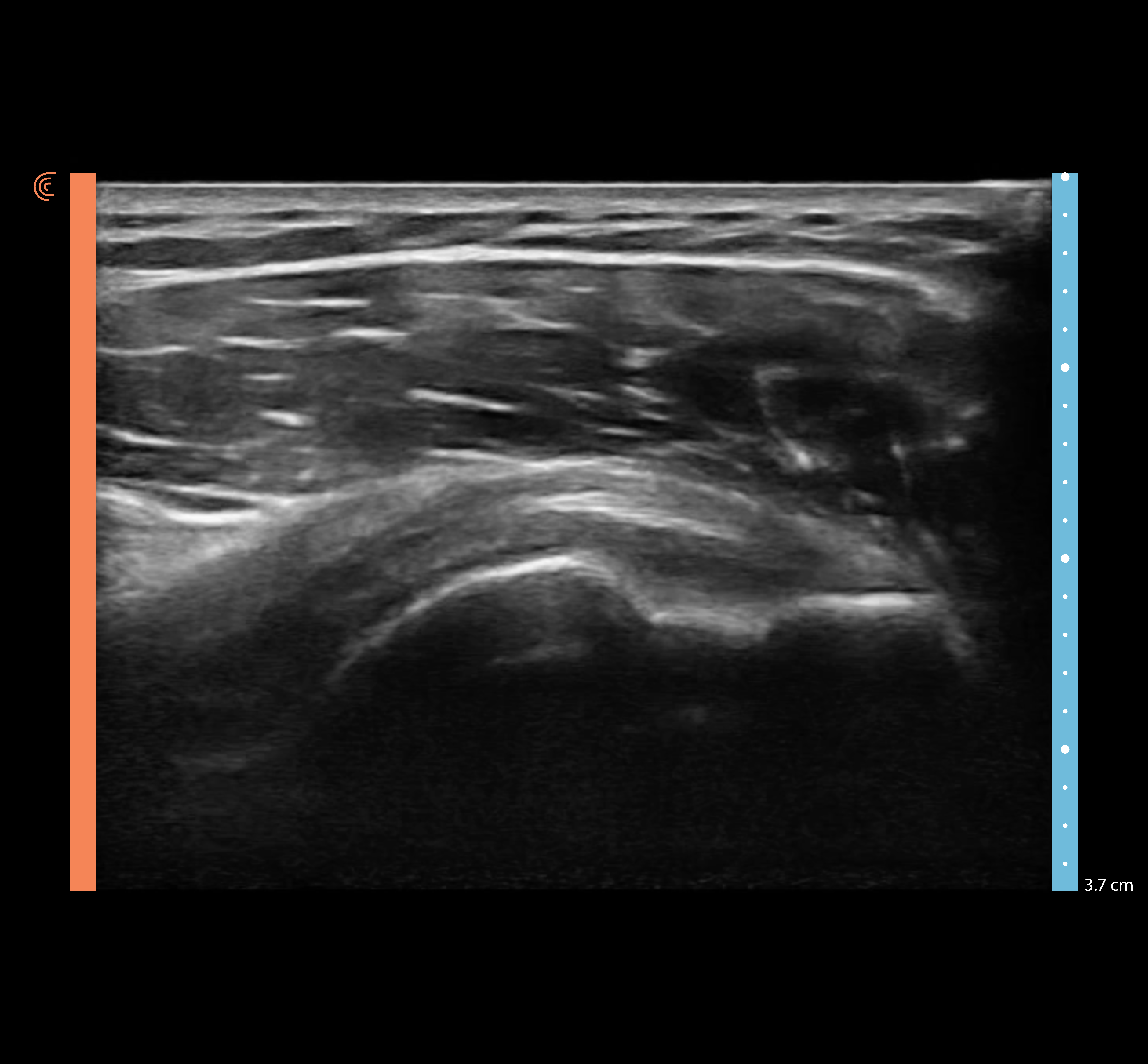

STEP 3 Subscapularis: Short Axis
Rotate the probe 90 degrees to visualize the tendon in short-axis view.
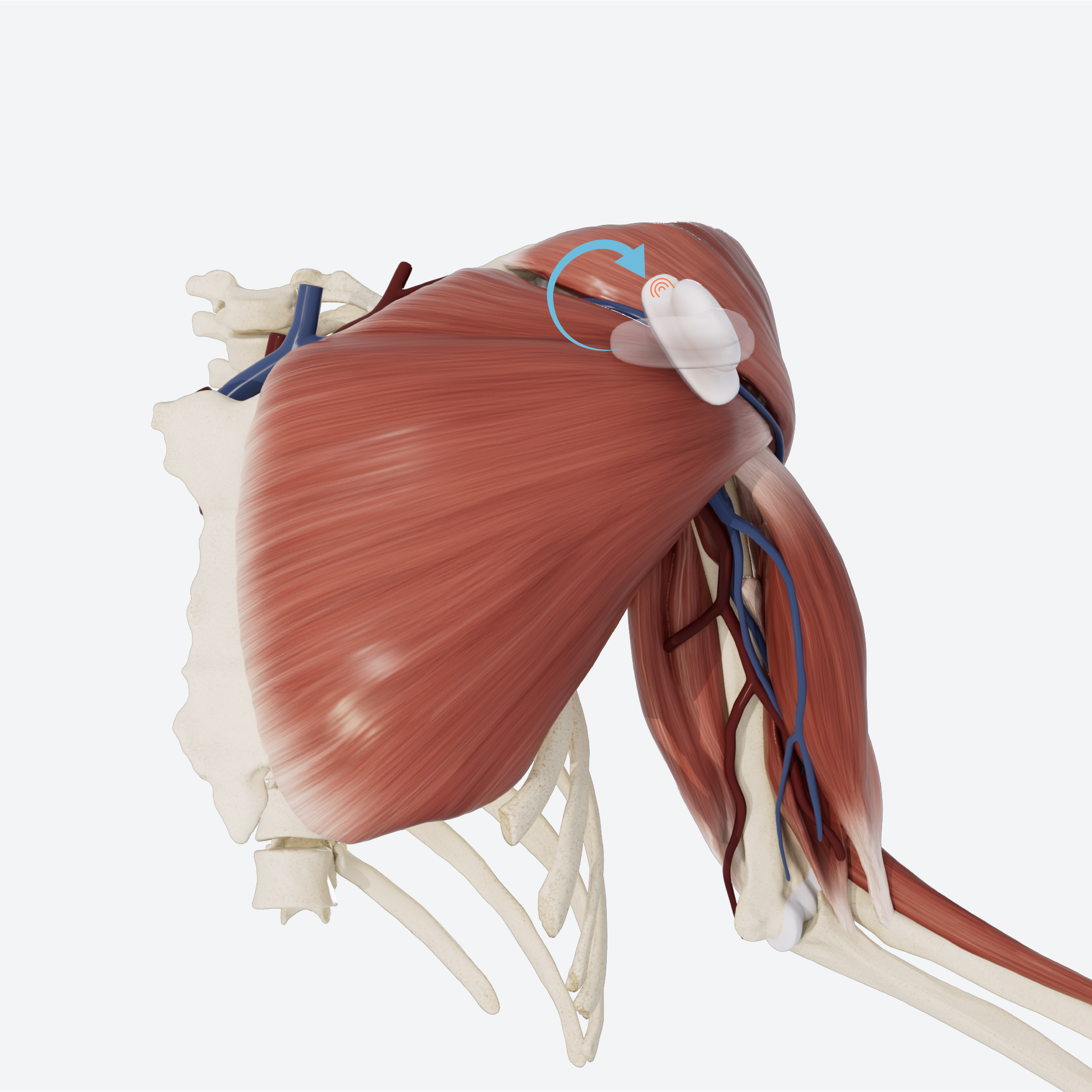
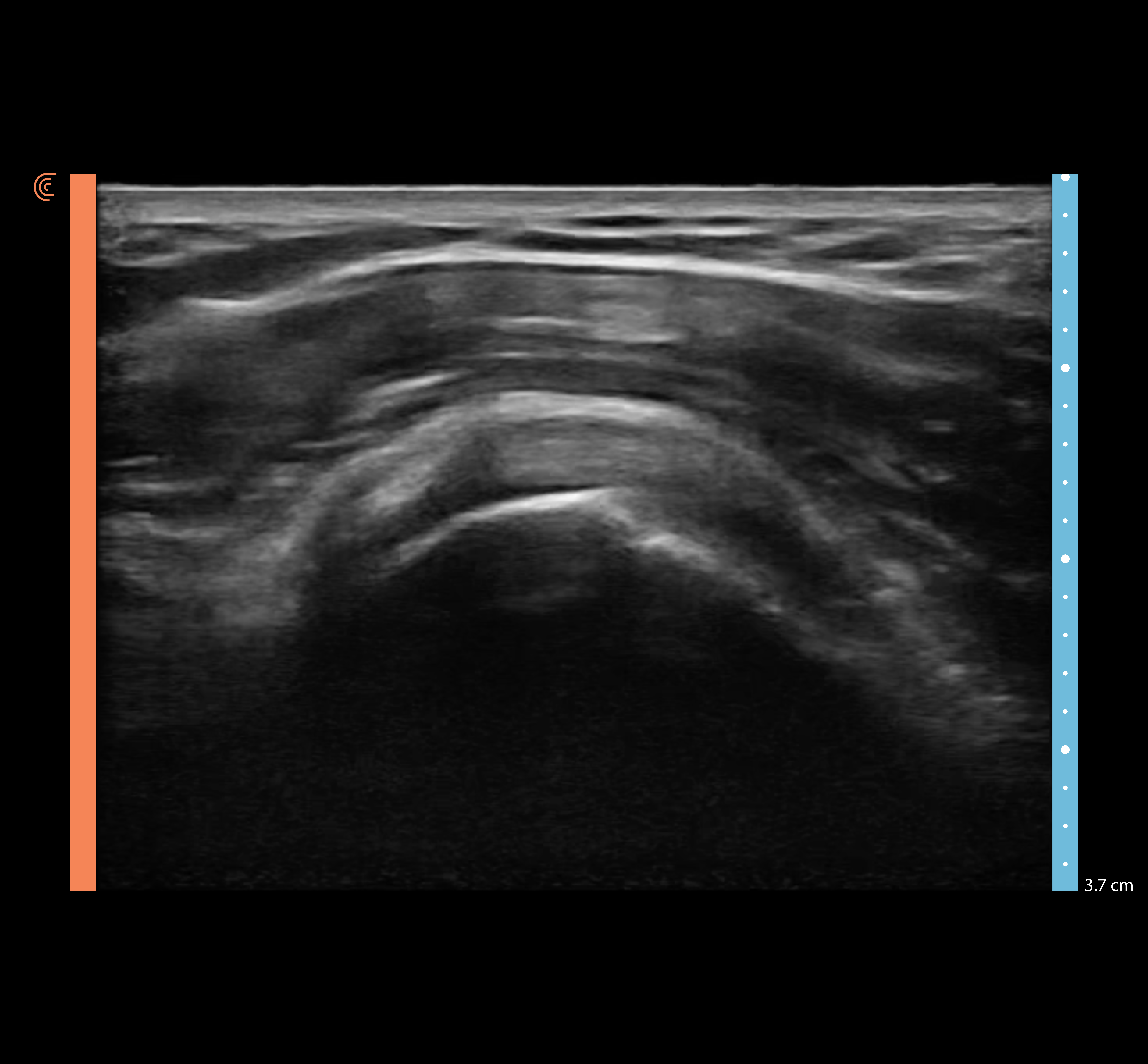

Note: The subscapularis tendon will appear fibrillar in a long-axis view, and heterogenous in a short-axis view. This is due to its characteristic fibular tendinous bundles.
REVIEW Anterior Transverse: Subscapularis
Use the slider to review the dynamic US technique shown through steps 1-3 to visualize subscapularis in external rotation.
