Module 2.1
Posterior Transverse: Infraspinatus and Glenohumeral Joint
In this section, we will use ultrasound to explore the shoulder from the posterior aspect. Structures visualized will include deltoid, infraspinatus, teres minor, the glenohumeral joint, and the glenoid labrum.
Patient starting position:
The patient’s arm should be relaxed and in neutral position.
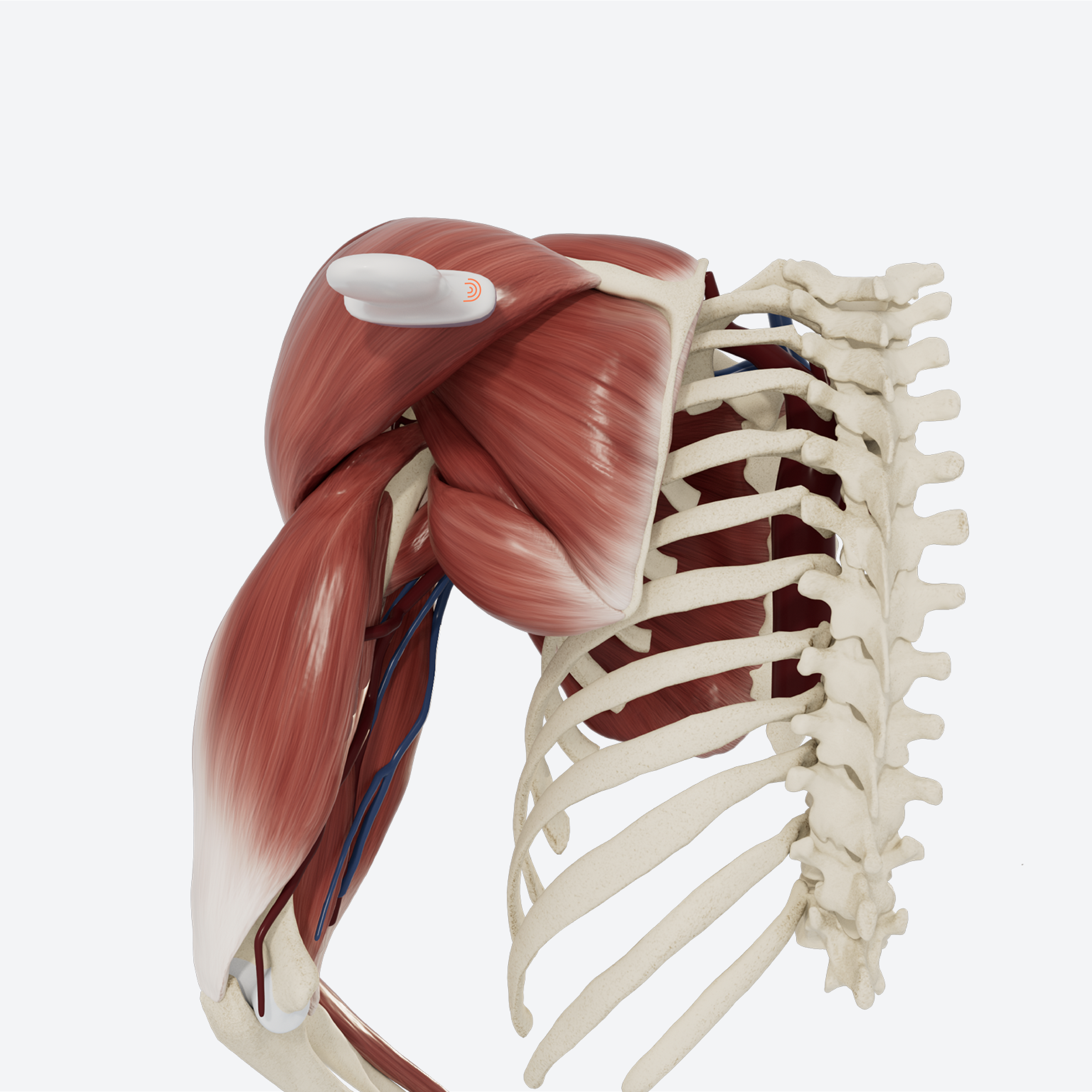
Note: this examination is demonstrated on the left shoulder, with the ultrasound probe indicator facing medially to begin. The numbers used to denote each step on the model correspond to the direction of the indicator. The orientation marker is always on the left side of the sonogram.
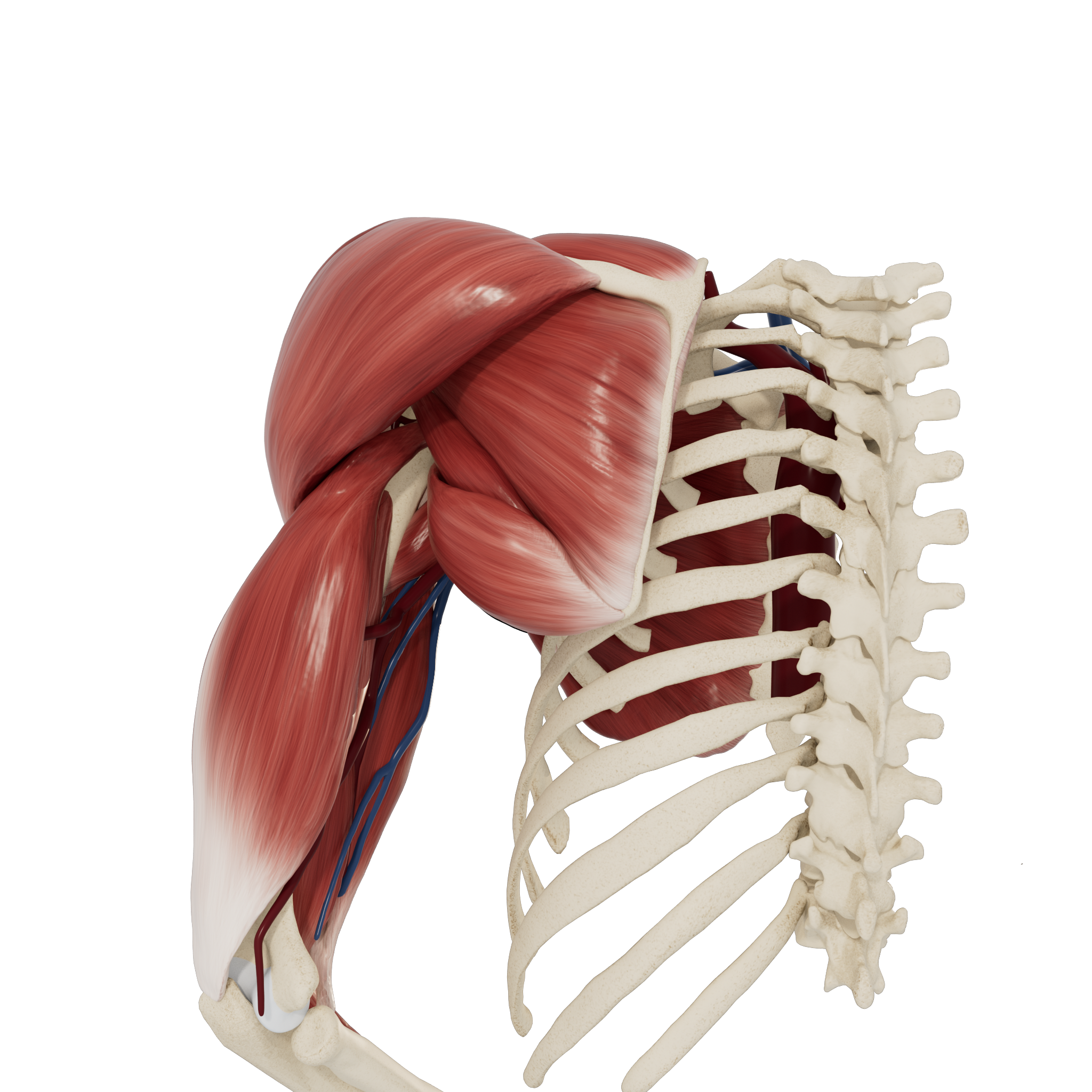
Patient starting position
STEP 1 Glenohumeral Joint
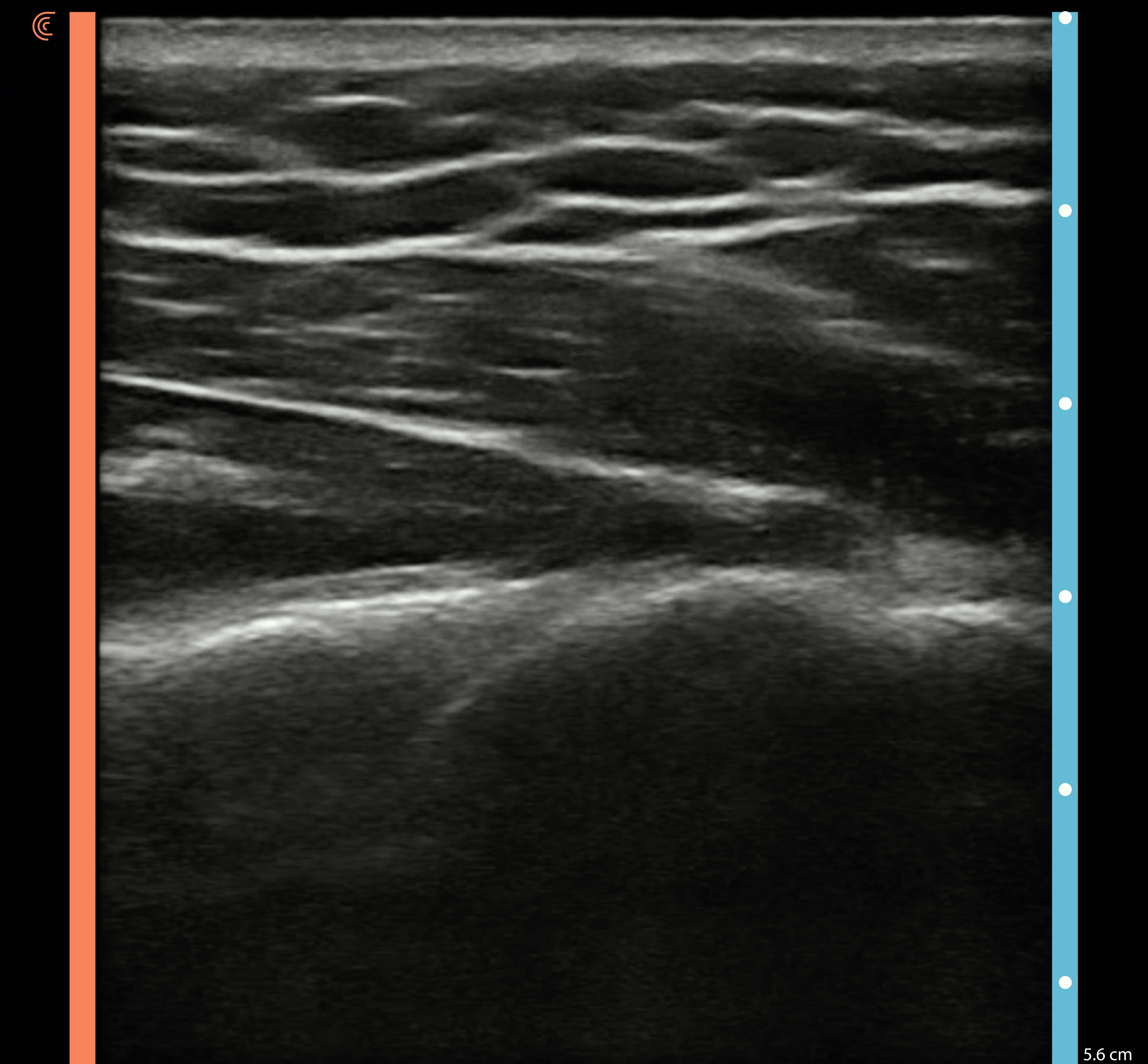
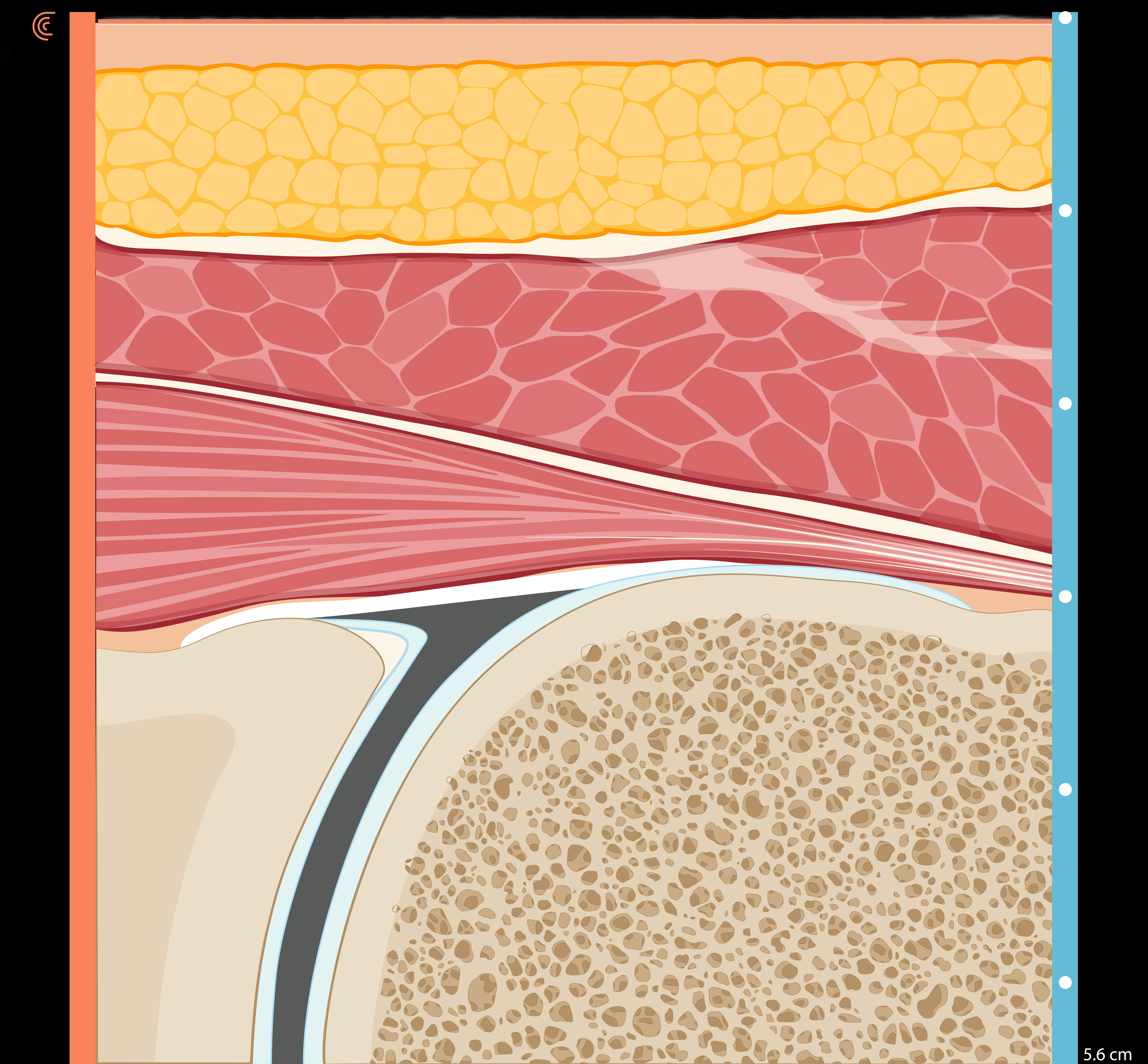
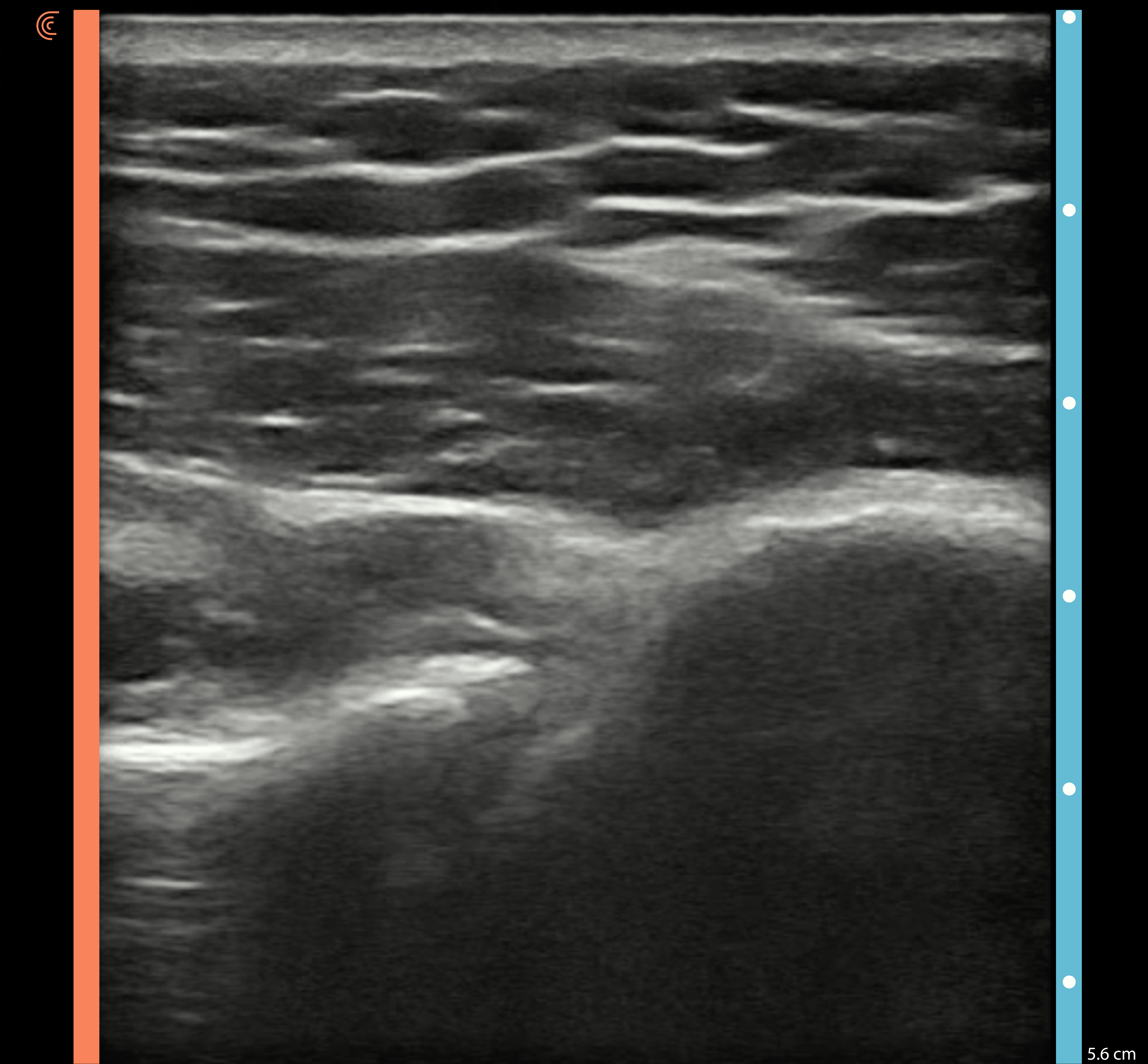
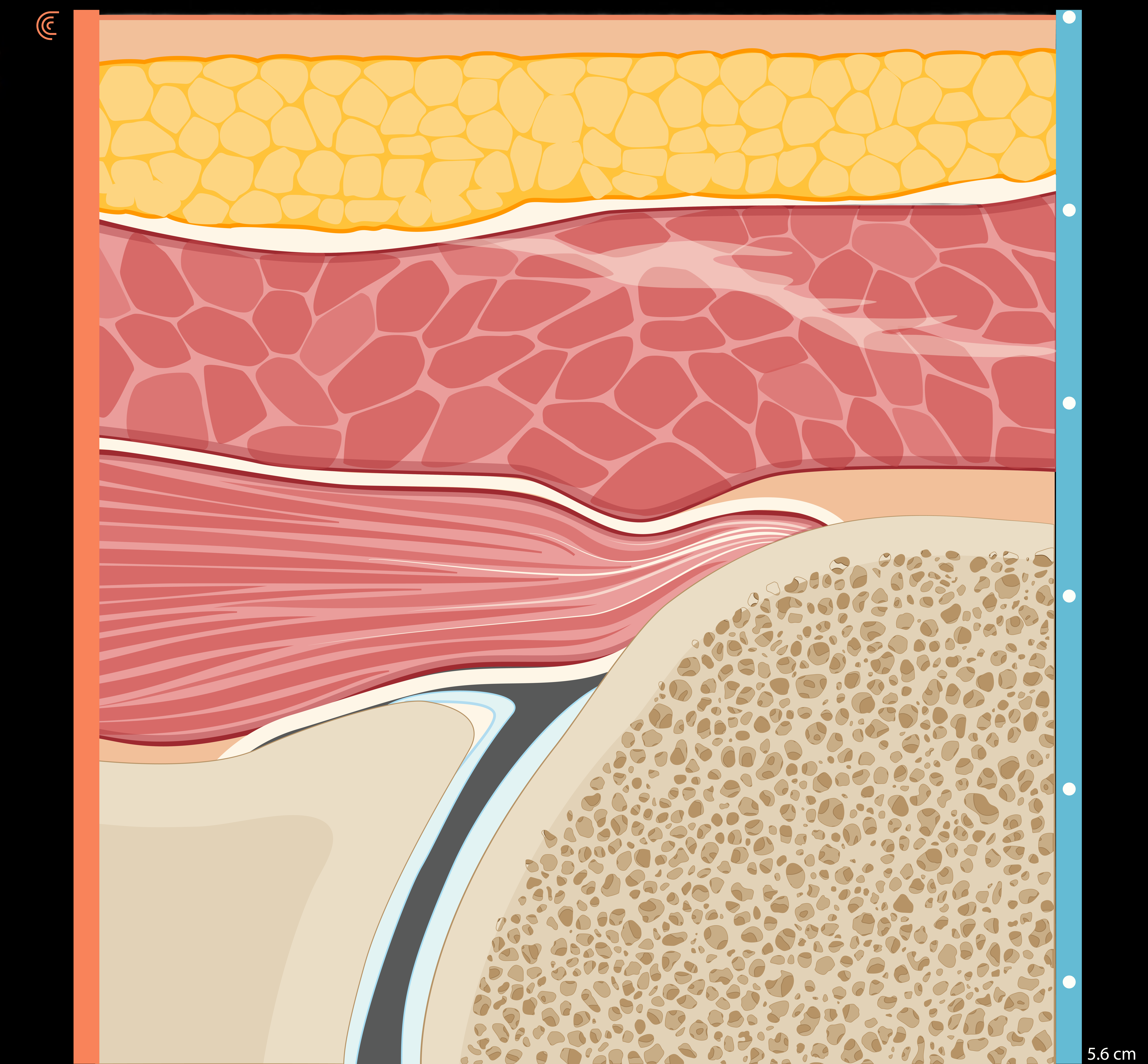
Begin with the probe oriented transversely directly below the scapular spine so that the posterior humeral head and glenoid fossa of the scapula are visualized. The synovial space maybe be observed as a thin hypoechoic line between the two hyperechoic bony structures. The glenoid labrum should appear as a triangle attached to the superficial aspect of the glenoid fossa, with an intermediate echolucency between the bone and the synovial space.
STEP 2 Glenohumeral Joint and Infraspinatus in Rotation
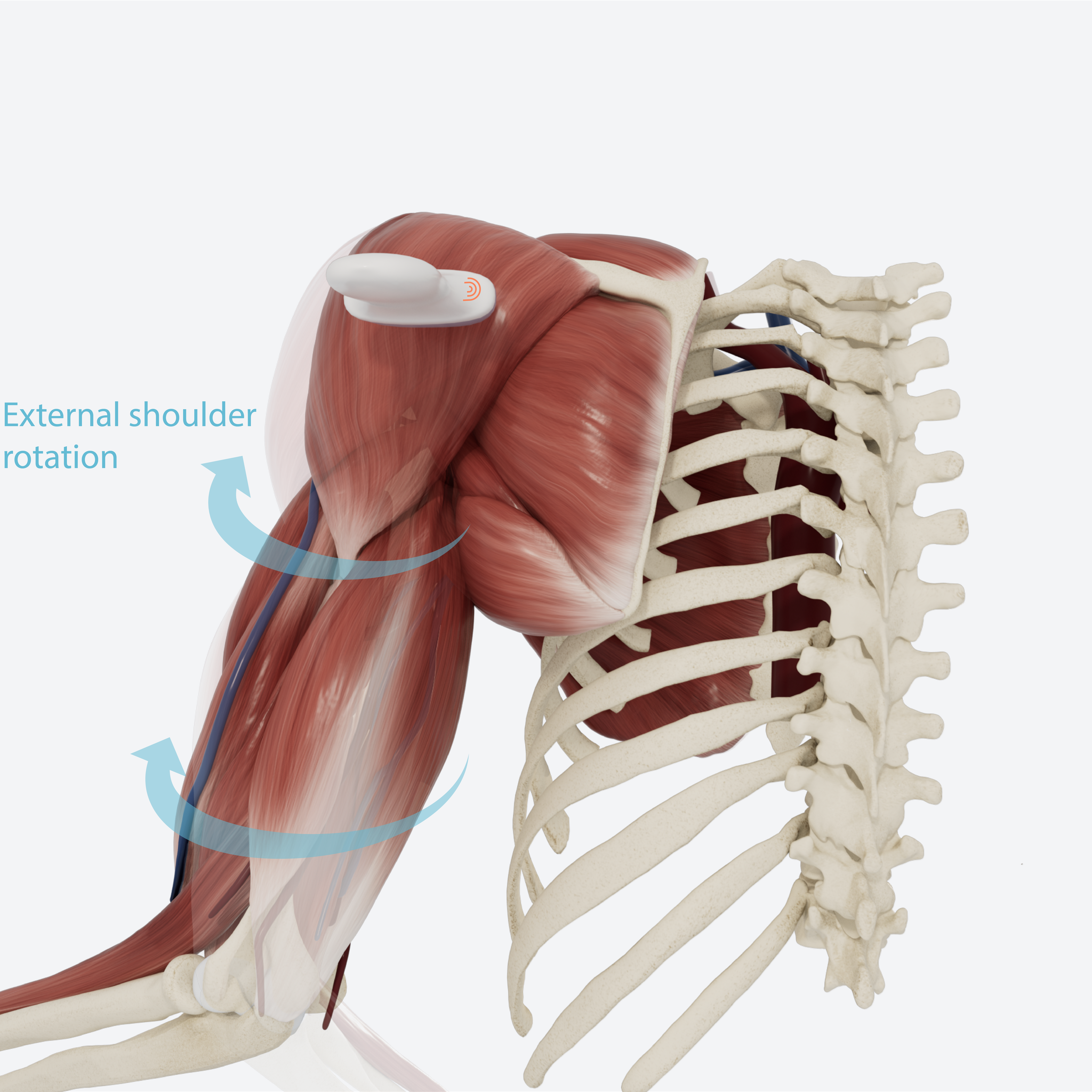
While keeping the probe still, have the patient externally rotate their arm to observe contraction of the infraspinatus pulling the greater tubercle toward the glenohumeral joint.
STEP 3 Infraspinatus: Long Axis
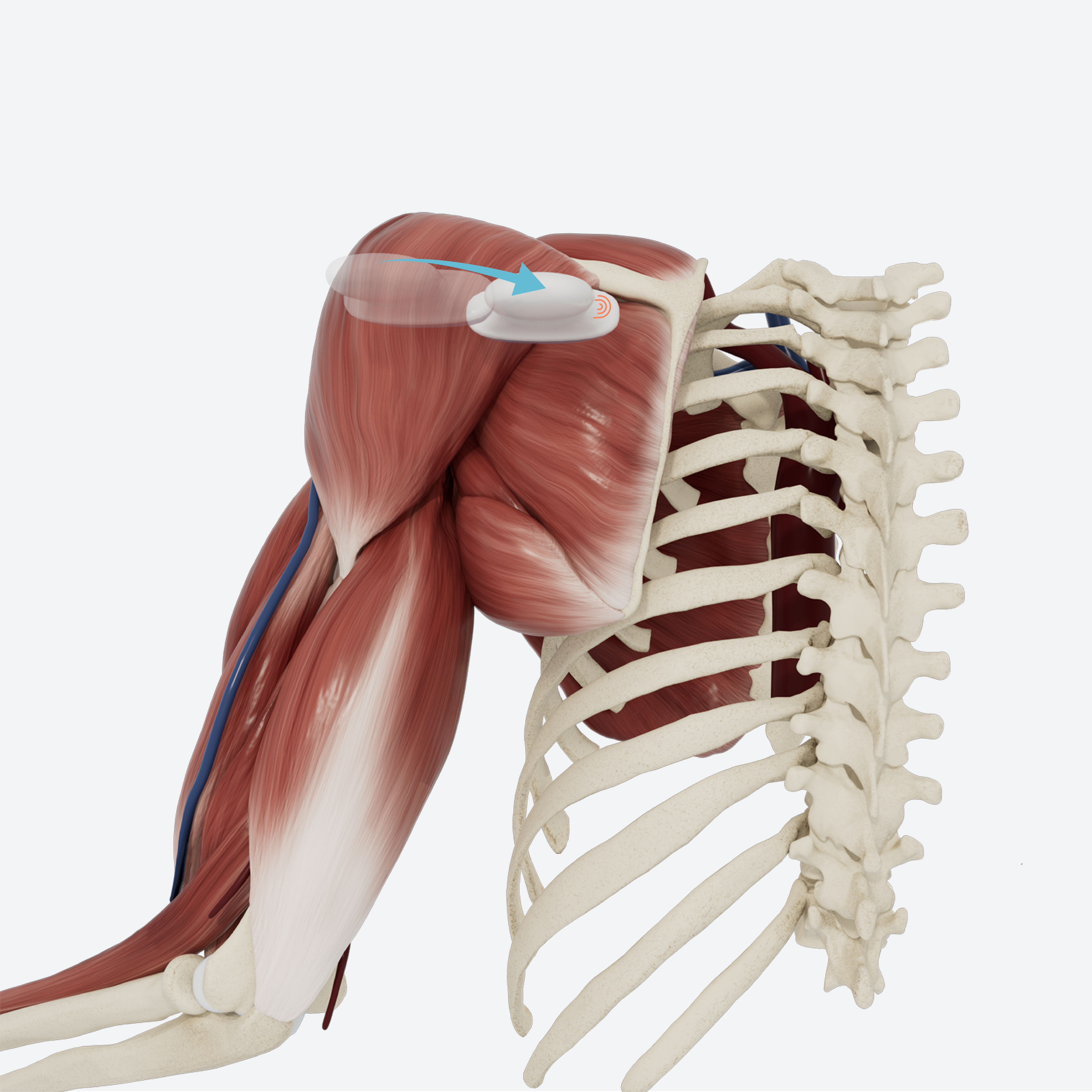
In the same view, superficial to the glenohumeral joint, the infraspinatus muscle can be observed in its long axis. Slide the transducer medially to view the muscles belly; slide laterally to view the tendon and its attachment to the greater tubercle.
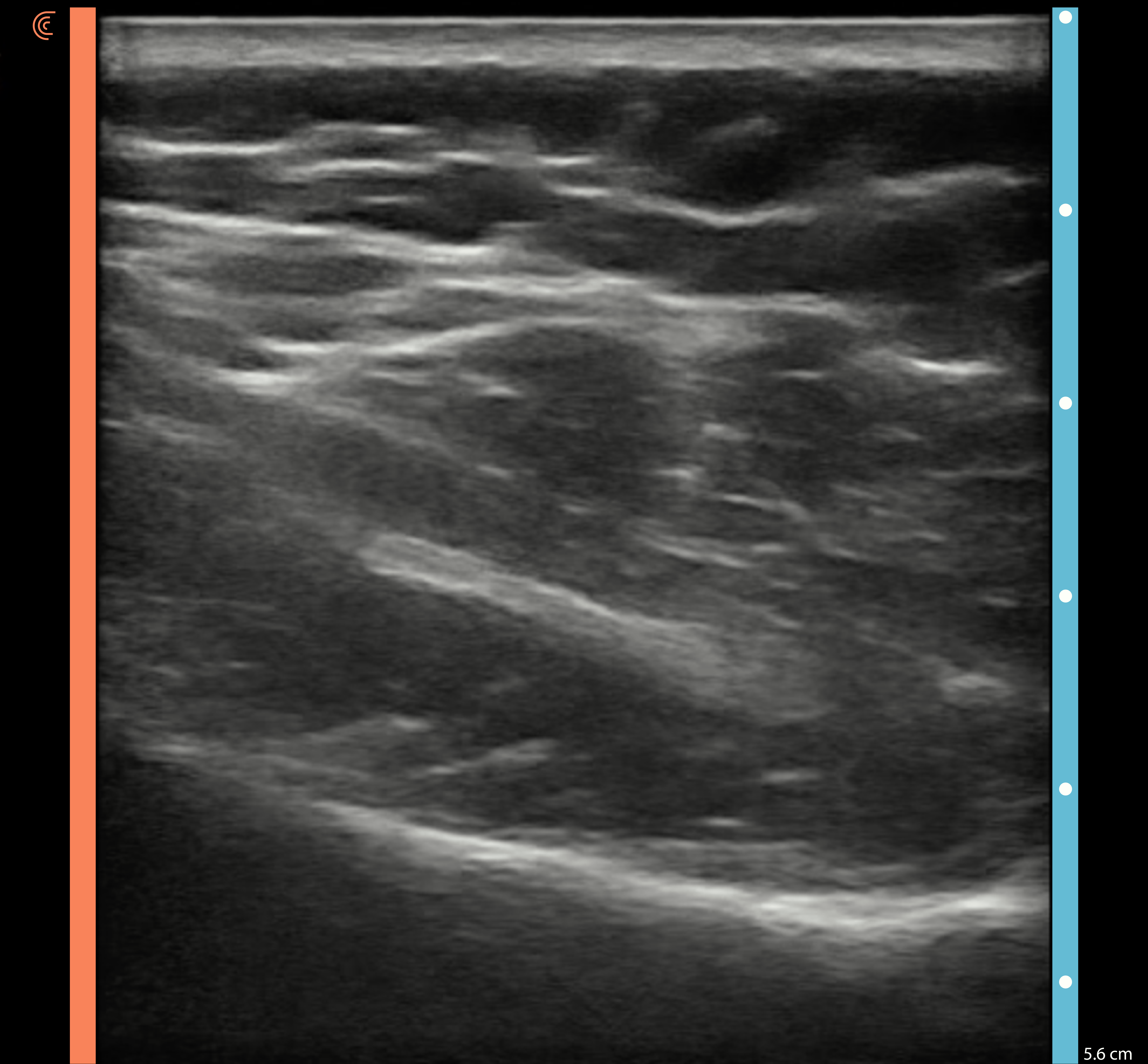

REVIEW Posterior Transverse: Infraspinatus and Glenohumeral Joint
Use the slider to review the dynamic US technique shown through steps 1-3 to visualize infraspinatus in external rotation.
