Module 3.1
Coronal Plane: Acromioclavicular Joint
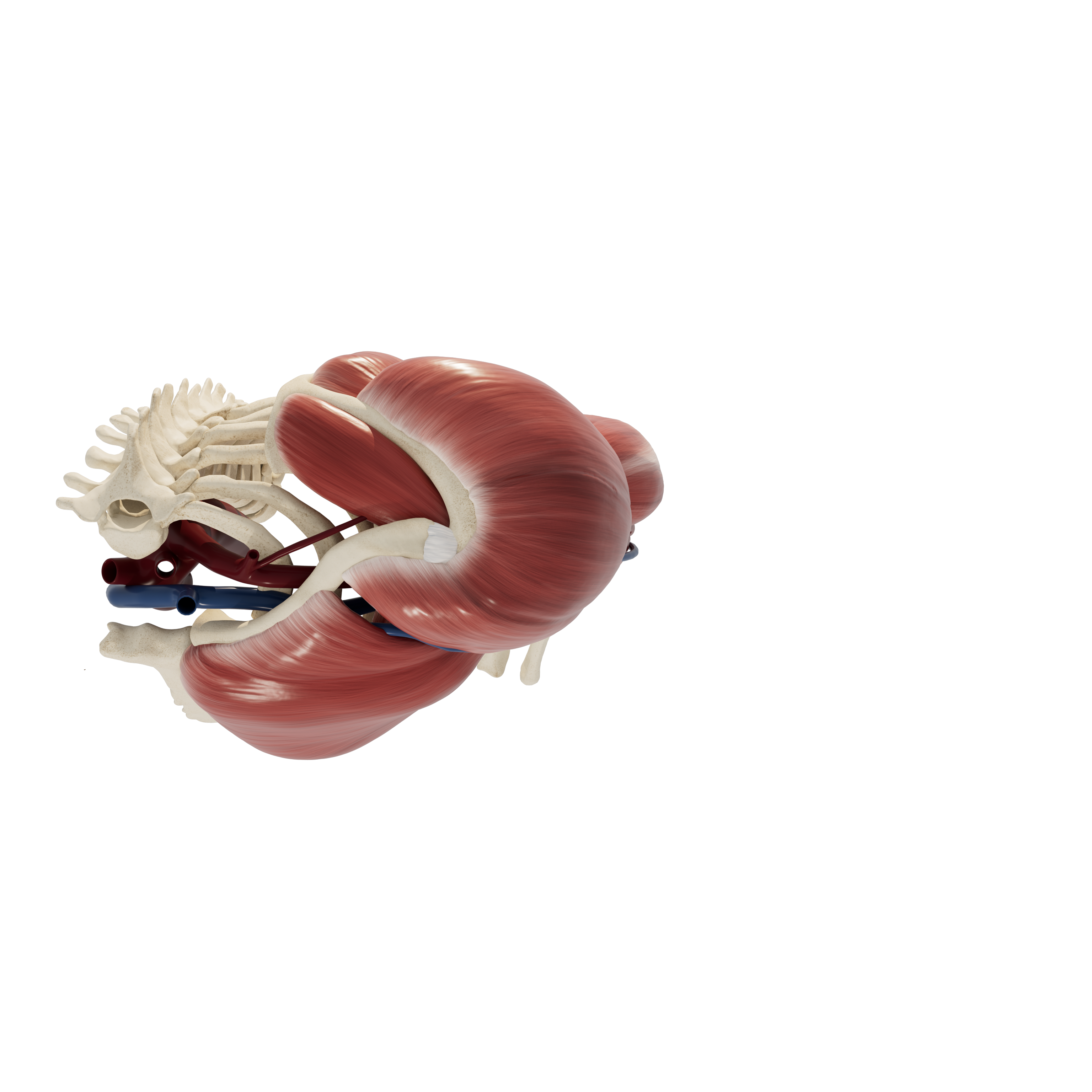
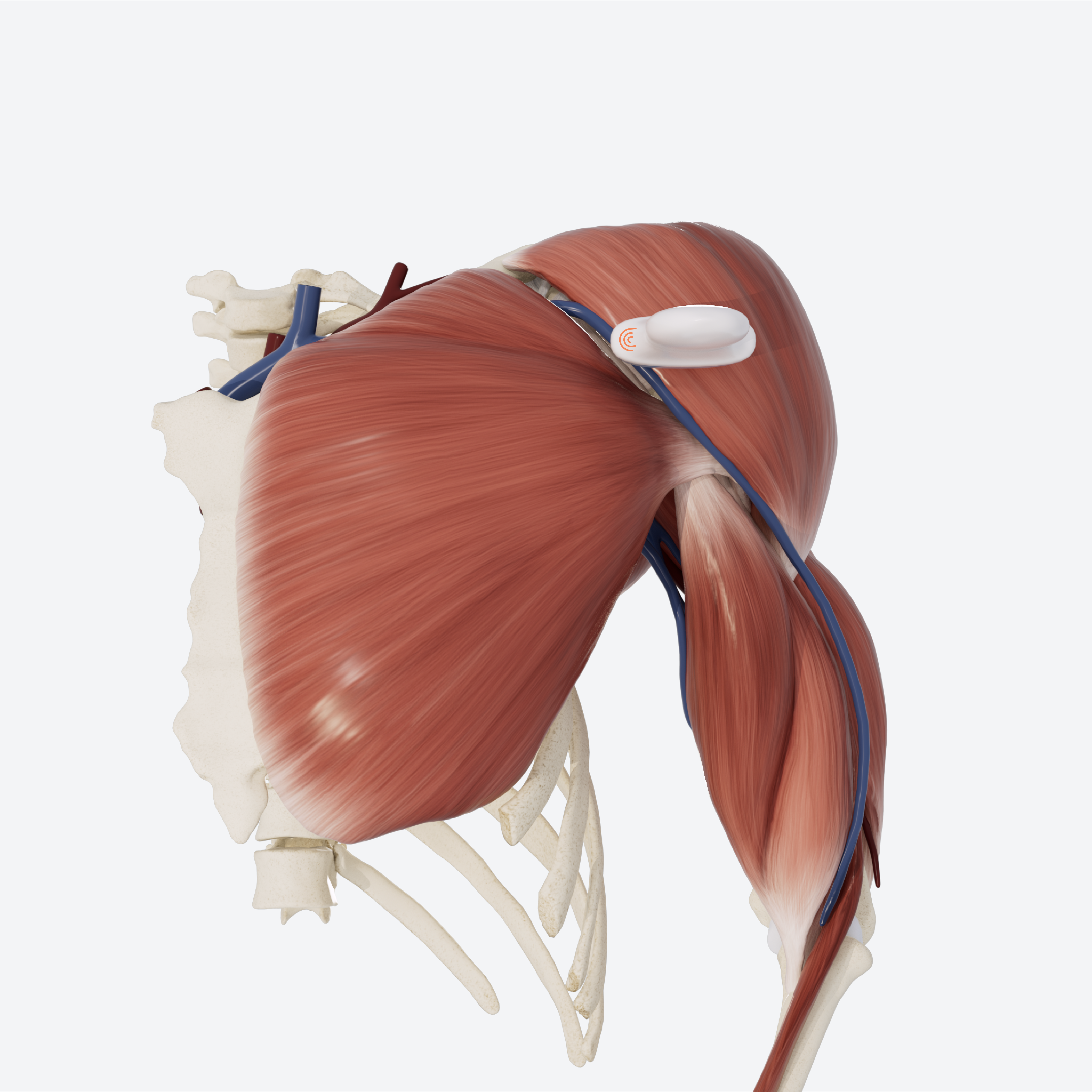
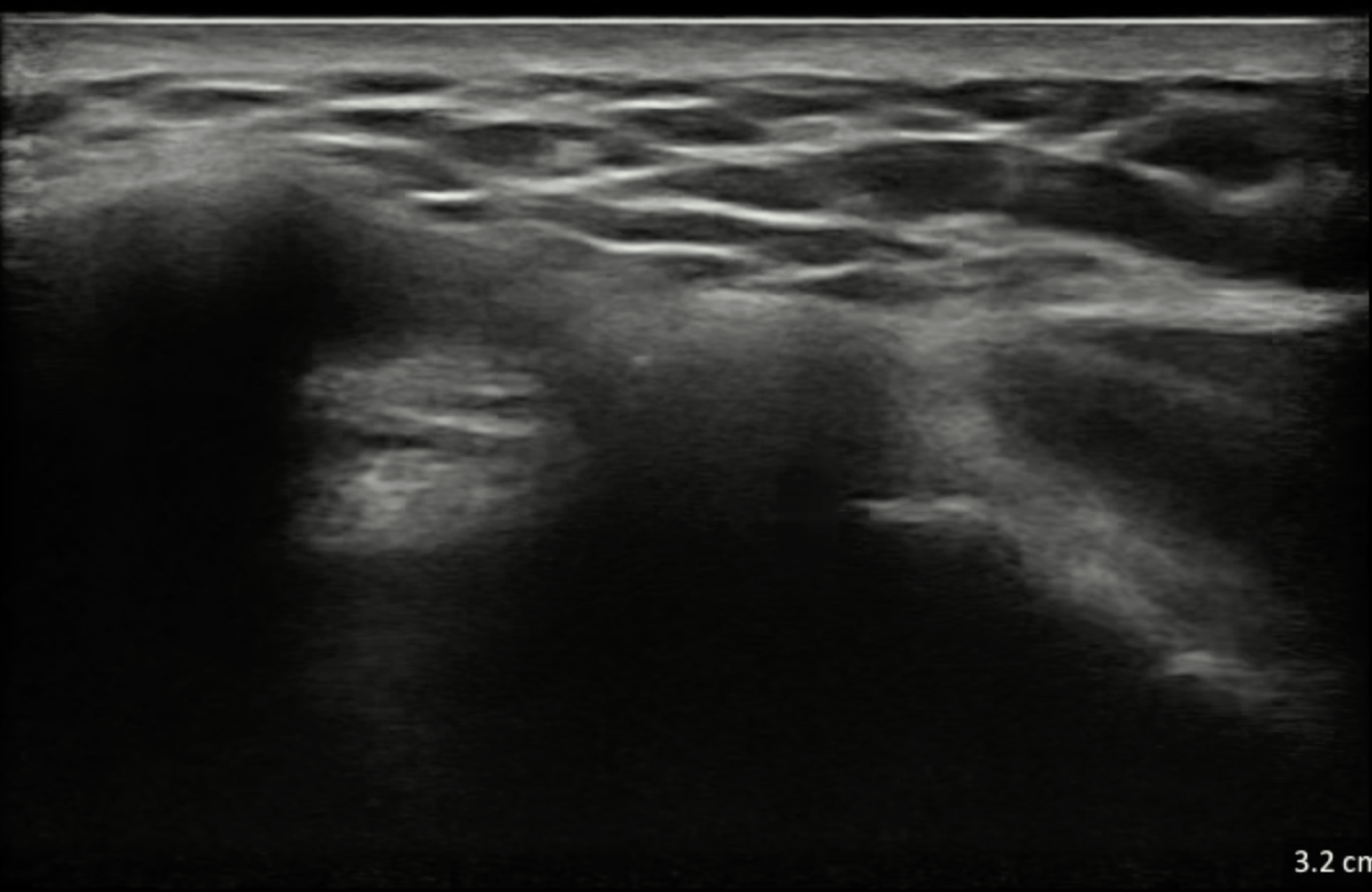
In this section, we will use ultrasound to explore the shoulder from the superior aspect in a coronal plane. Structures visualized will include the acromion, aromioclavicular joint and ligament, and supraspinatus muscle.
Patient starting position:
The patient’s arm should be relaxed and in neutral position.
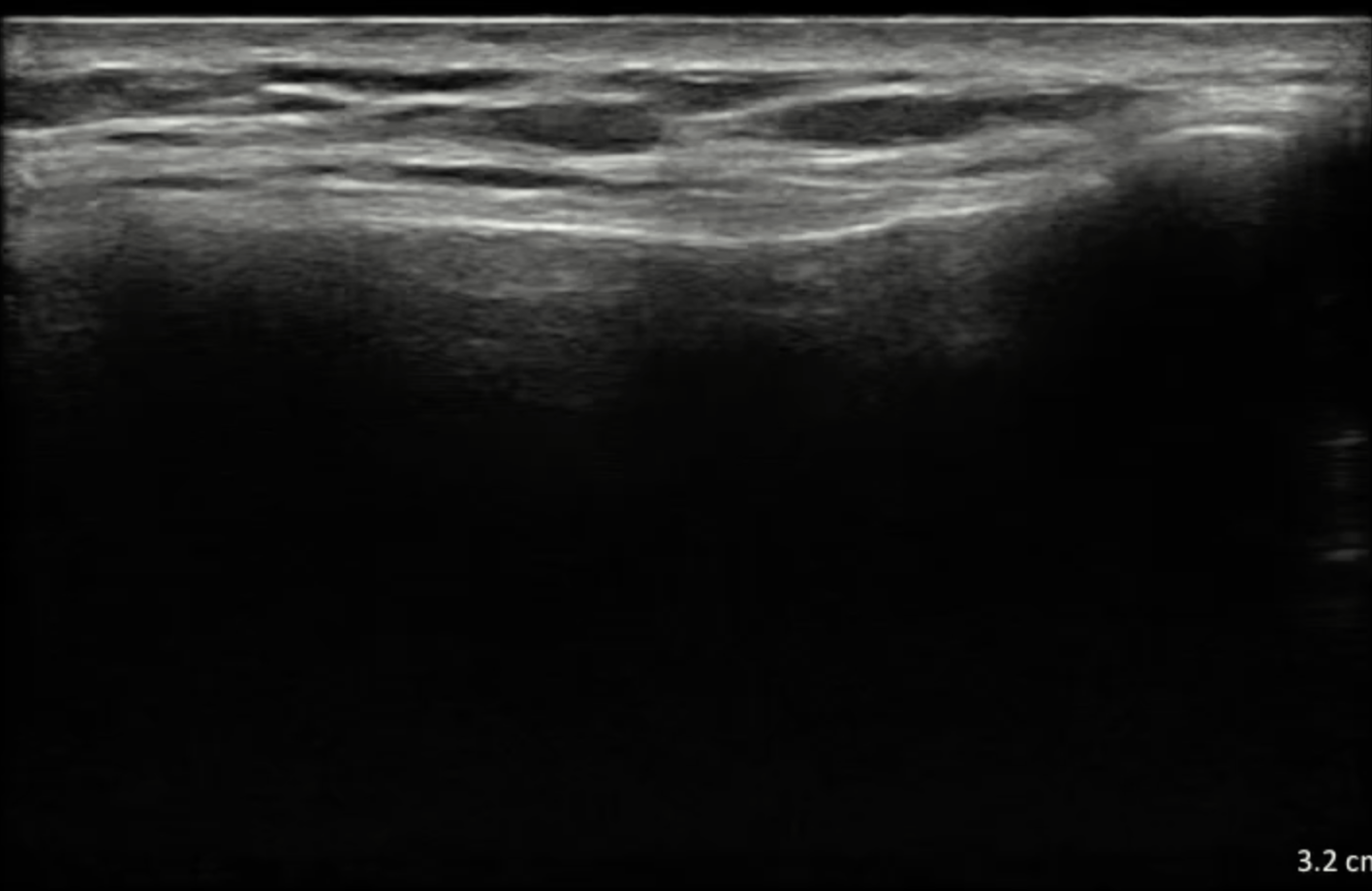
Note: this examination is demonstrated on the left shoulder, with the ultrasound probe indicator facing medially to begin. The numbers used to denote each step on the model correspond to the direction of the indicator. The orientation marker is always on the left side of the sonogram.
Patient starting position
STEP 1 Acromioclavicular Joint
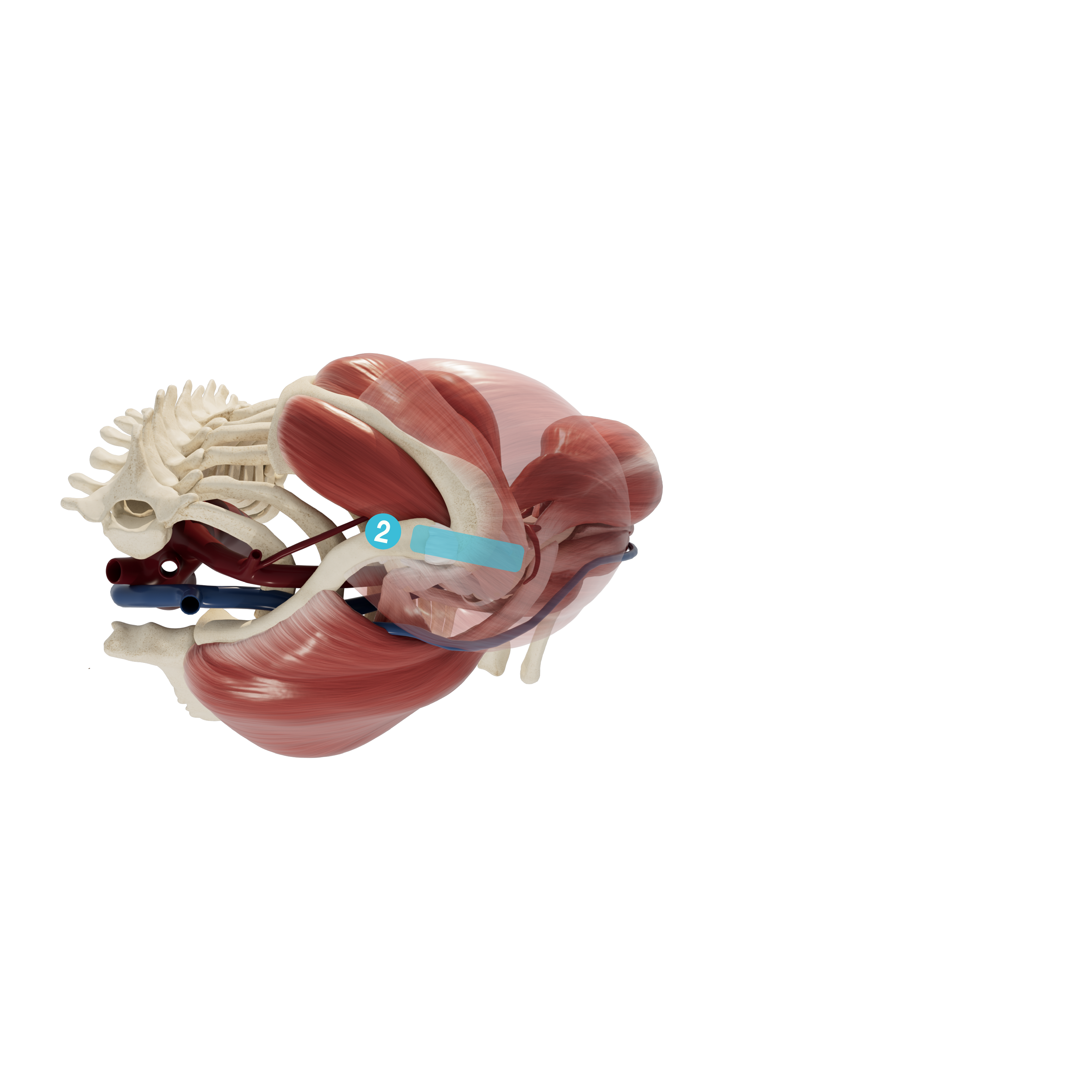
Begin by placing the transducer directly on top of the clavicle, in a coronal plane. The hyperechoic surface of the clavicle can be observed very superficially. Slide the transducer laterally on the clavicle until a hypoechoic v-shaped “gap” between the bones is observed. This is the acromioclavicular joint. Identify the distal end of the clavicle medial to the join, the acromion lateral to the joint, and ligaments superficial to the joint.

STEP 2 Acromion and Supraspinatus
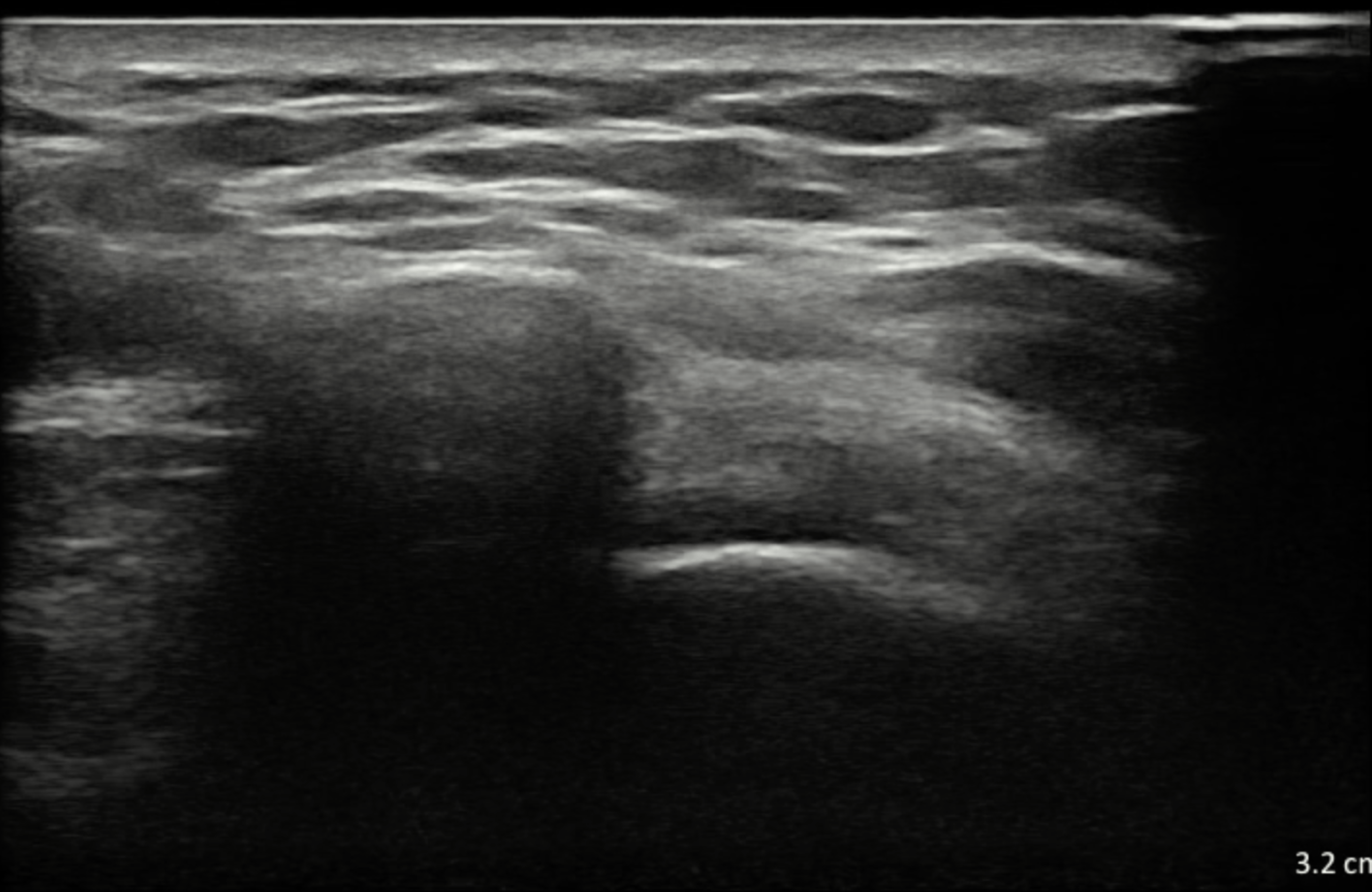
While keeping the probe still, have the patient externally rotate their arm with their forearm slightly supinated and their elbow anchored against the torso. This will stretch subscapularis into view. Slide the probe superiorly and inferiorly on the tendon to view insertion width on the lesser tubercle.

STEP 2 Supraspinatus in Shoulder Abduction
While keeping the probe still, have the patient abduct their shoulder to 90 degrees with the arm in internal rotation. You should be able to watch as the supraspinatus pulls the greater tubercle into the subacromial space.

REVIEW Anterior Transverse: Biceps Tendon
Use the slider to review the dynamic US technique shown through steps 1-4 to visualize the entire biceps tendon.
