Module 3.2
Superior Modified Crass: Supraspinatus
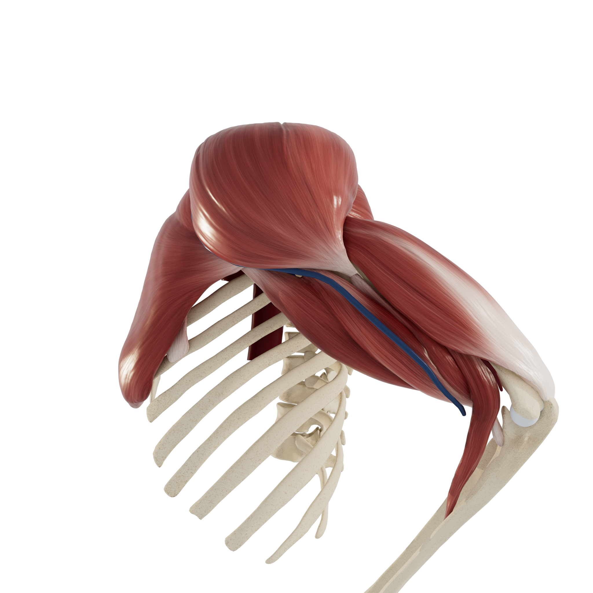
In this section, we will use ultrasound to explore supraspinatus muscle and tendon in the modified crass position. Structures visualized will include the supraspinatus and infraspinatus muscles, biceps tendon, acromion, greater tubercle, and subacromial subdeltoid bursa.
Patient starting position:
Place the patient’s palm on the posterior aspect of their ipsilateral hip (iliac crest), so that their elbow is flexed and directed posteriorly. This is the Modified Crass position. This position brings most of the supraspinatus muscle out from under the acromion to allow ultrasound examination
Note: this examination is demonstrated on the left shoulder, with the ultrasound probe indicator facing medially to begin. The numbers used to denote each step on the model correspond to the direction of the indicator. The orientation marker is always on the left side of the sonogram.

Patient starting position
STEP 1 Supraspinatus Muscle Belly: Long Axis
Place the transducer parallel and superior to the spine of the scapula, pointing inferiorly. The bulk of the supraspinatus muscle belly can be visualized in long axis, passing laterally under the acromion.


STEP 2 Supraspinatus Tendon: Long Axis
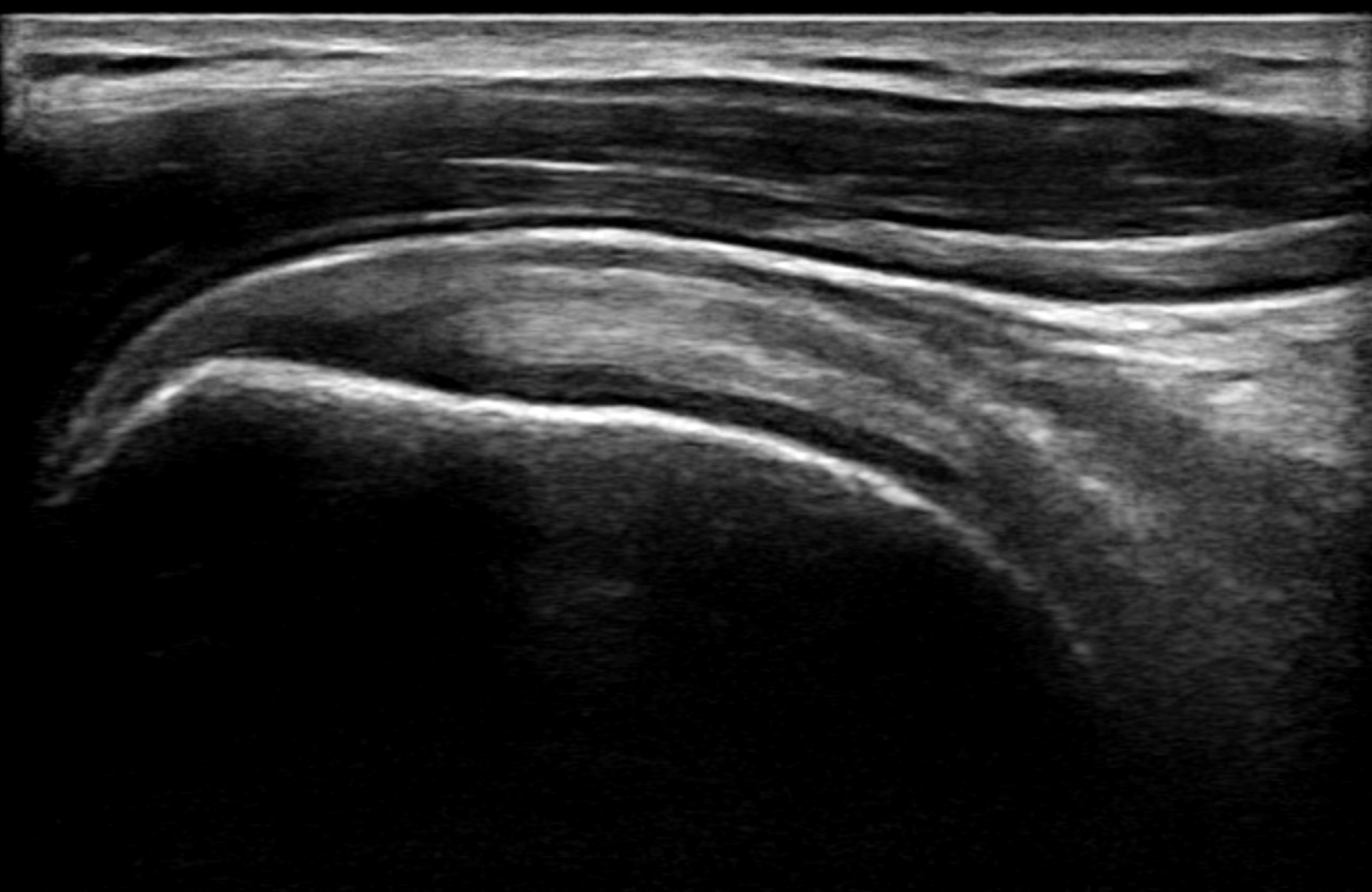
Slide the transducer laterally across the acromion. Observe the supraspinatus muscle re-emerging on the lateral side of the acromion and transitioning into its tendonous portion. Rock the transducer to follow the contour of the tip of the shoulder. Visualize the insertion of supraspinatus onto the greater tubercle. Note the thin hypoechoic subacromial subdeltoid bursa directly superficial to the supraspinatus.


STEP 3 Supraspinatus Tendon: Short Axis
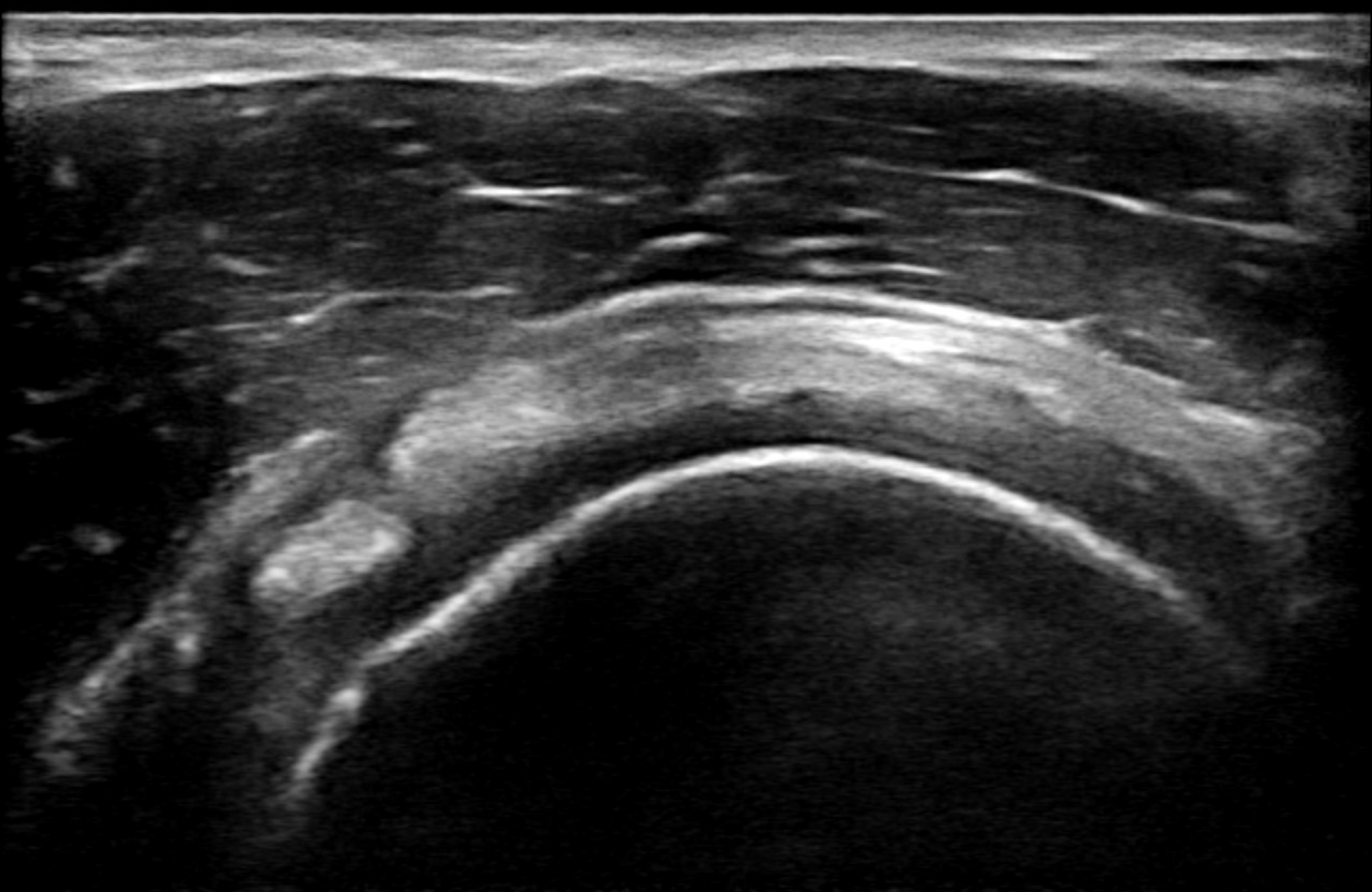
Rotate the transducer 90 degrees on the tip of the shoulder to visualize the supraspinatus tendon in its short axis. Observe the hyperechoic long head of the biceps tendon in its short axis forming the medial border of the supraspinatus. The infraspinatus tendon can be seen posterior to the supraspinatus with obliquely running fibres.


REVIEW Anterior Transverse: Biceps Tendon
Use the slider to review the dynamic US technique shown through steps 1-4 to visualize the entire biceps tendon.
