Module 2.2
Posterior Sagittal: Infraspinatus and Teres Minor
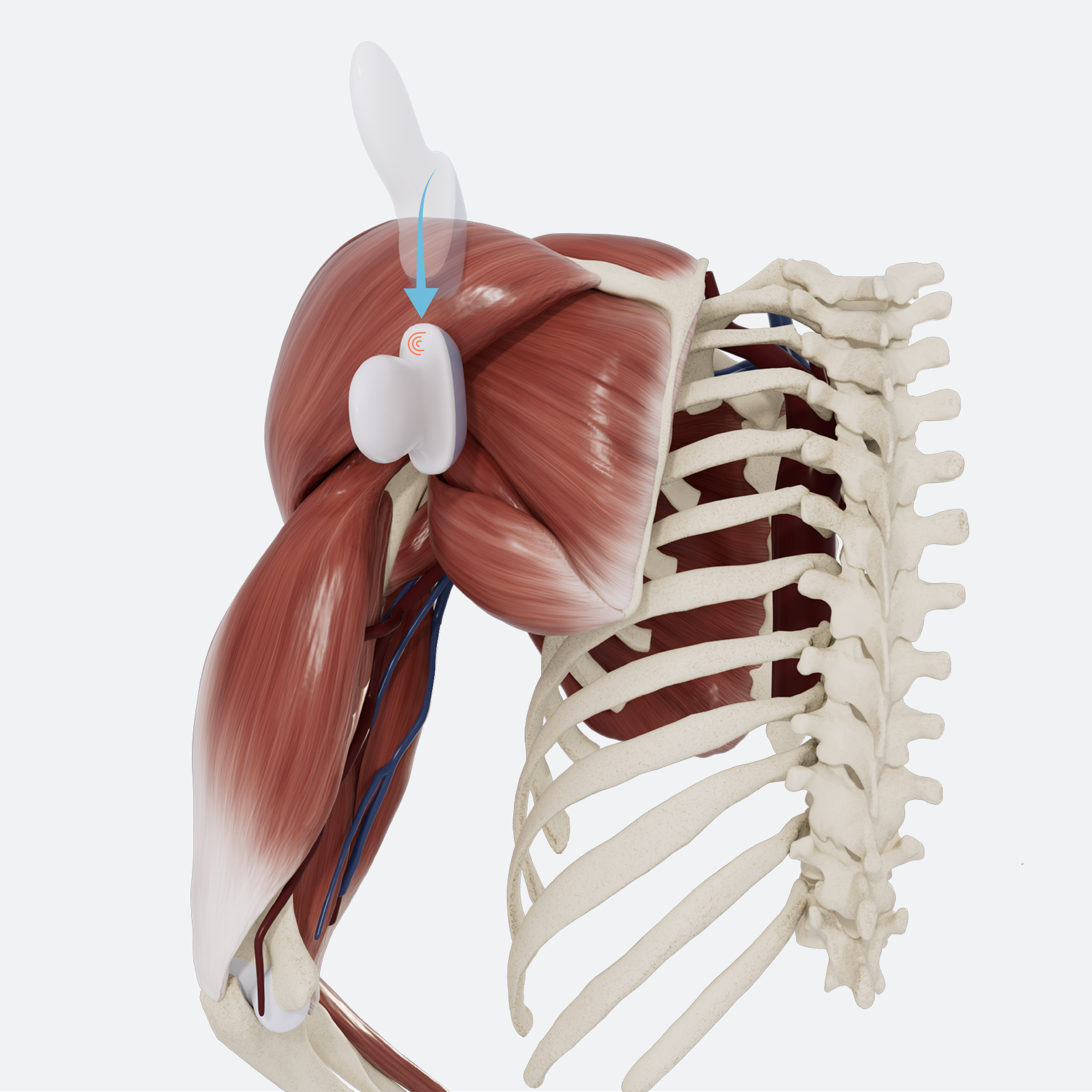
In this section, we will use ultrasound to explore the spatial relationship between infraspinatus and teres minor, and their attachments on the scapula. Structures visualized will include the spine of the scapula, the posterior deltoid, infraspinatus, and teres minor.
Patient starting position:
The patient’s arm should be relaxed and in neutral position.
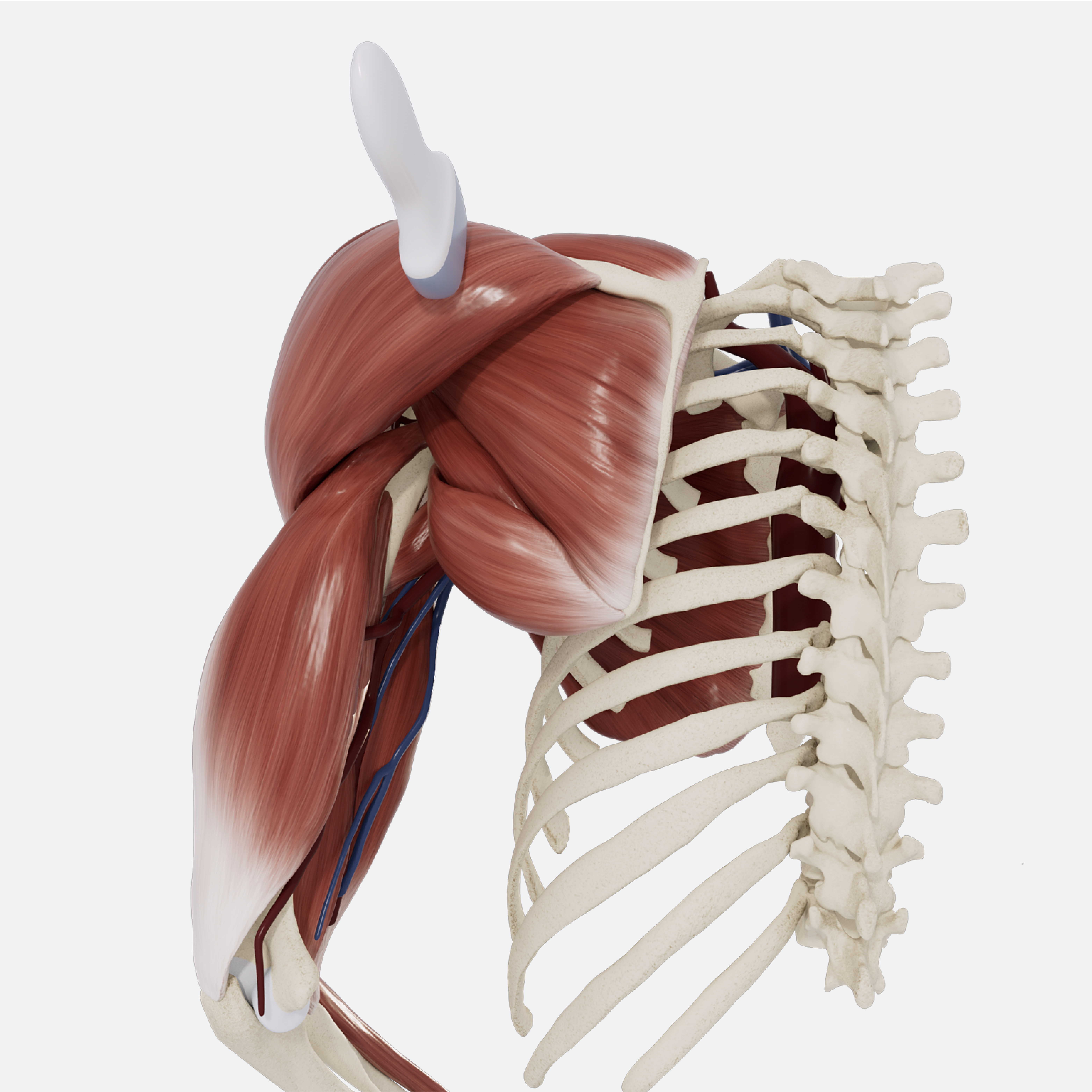
Note: this examination is demonstrated on the left shoulder, with the ultrasound probe indicator facing superiorly to begin. The numbers used to denote each step on the model correspond to the direction of the indicator. The orientation marker is always on the left side of the sonogram.
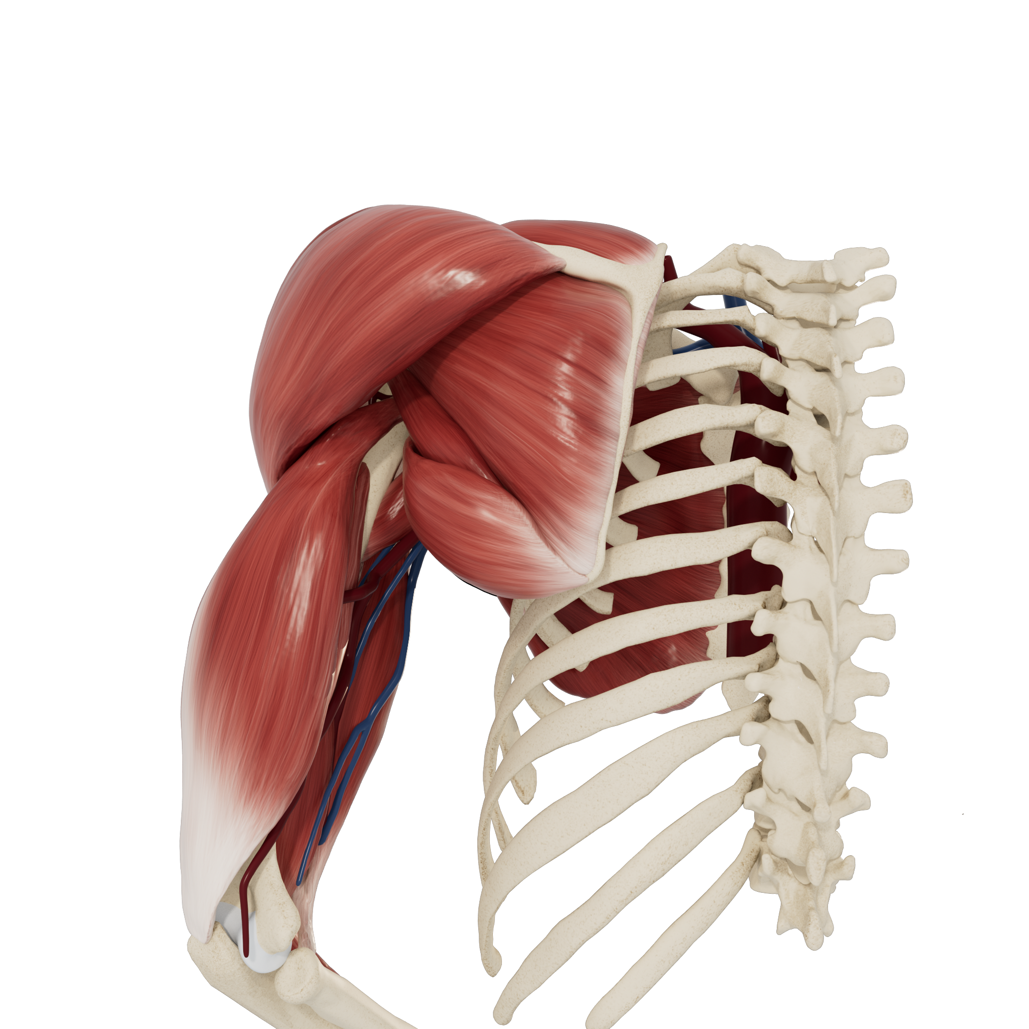
Patient starting position
STEP 1 Infraspinatus: Short Axis
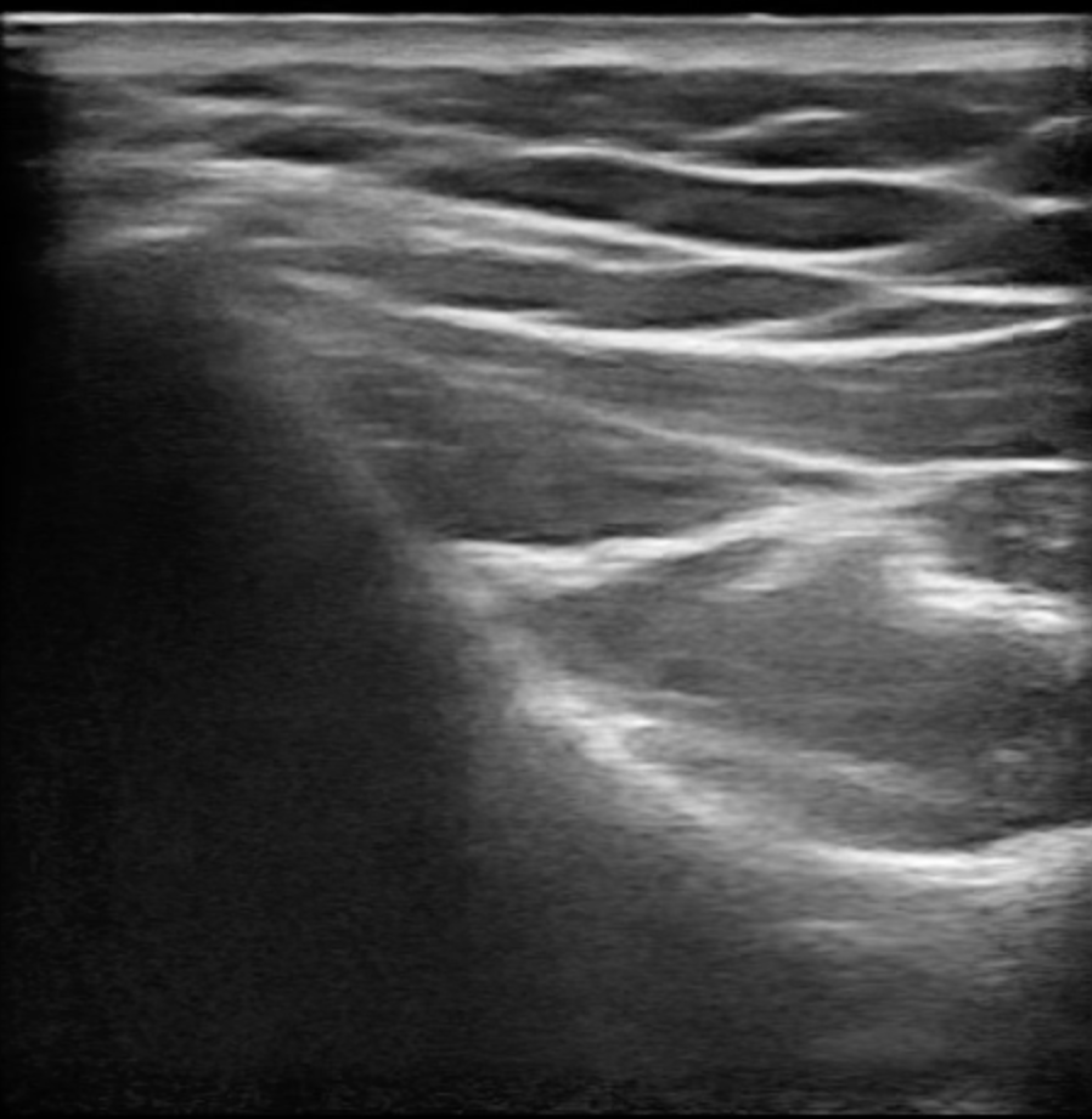
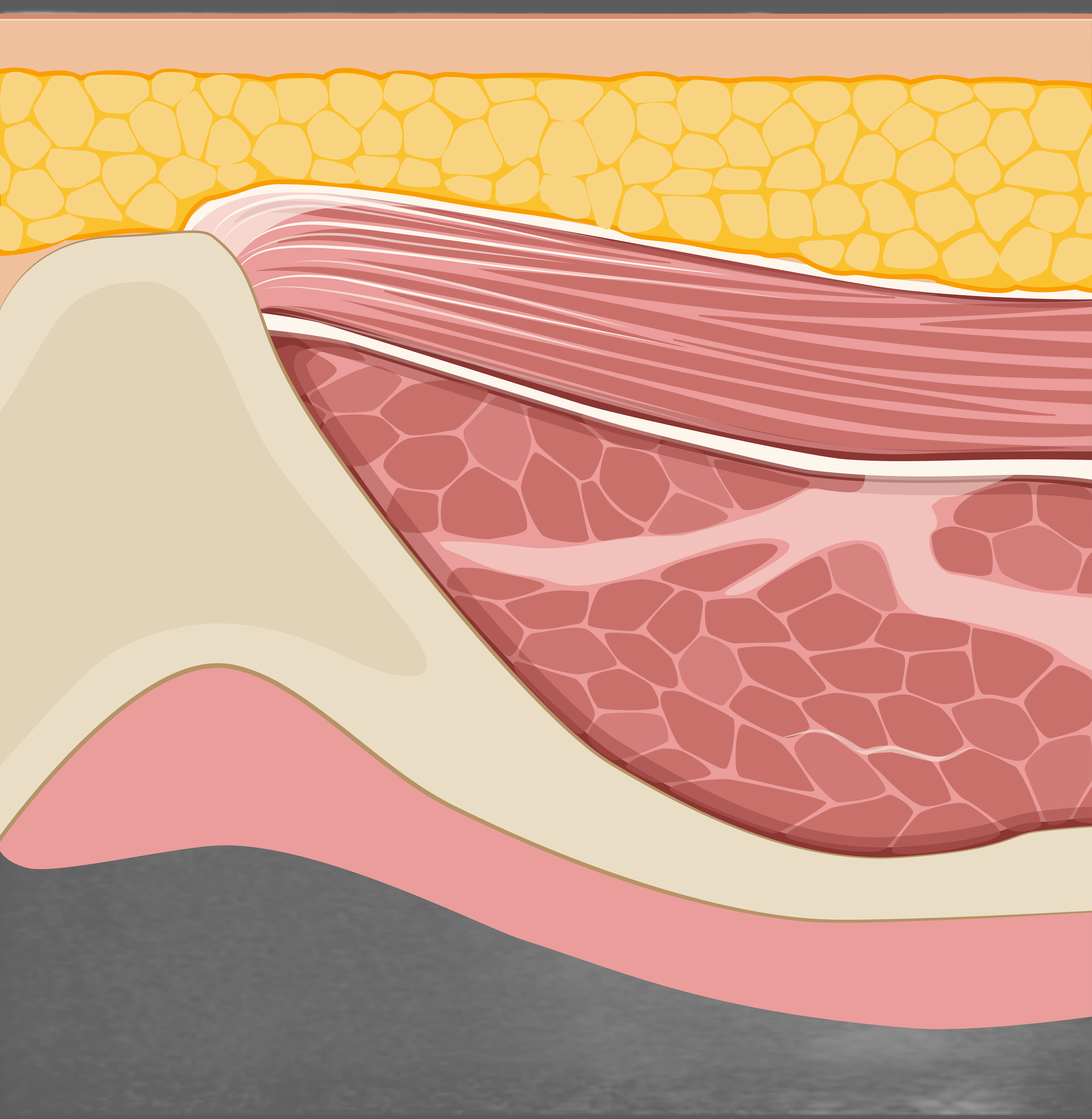
Begin with the transducer in the sagittal plane directly below the scapular spine and just medial to the acromion. The infraspinatus muscle belly in short axis can be observed directly against the scapula.
STEP 2 Teres Minor: Short Axis
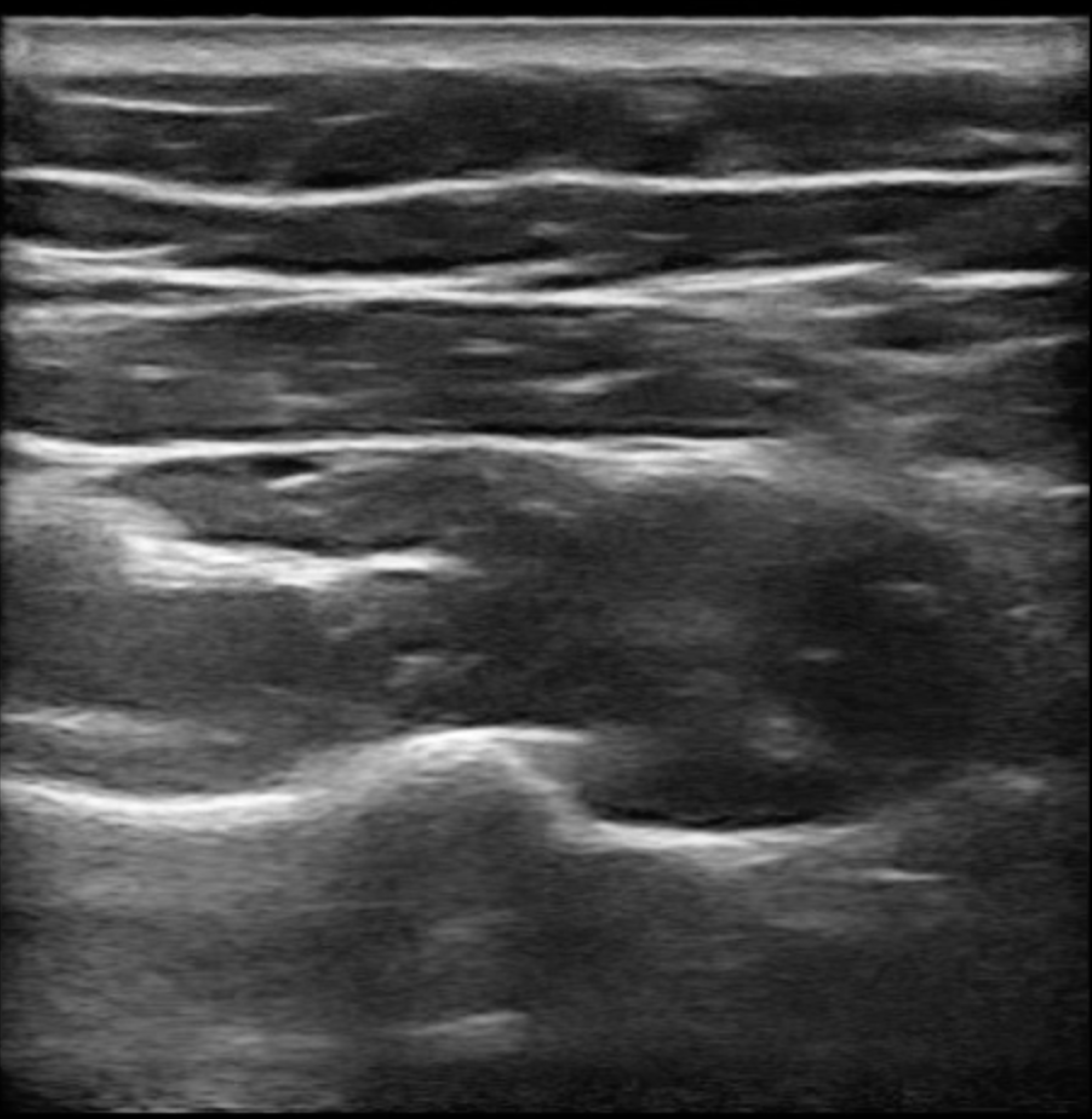
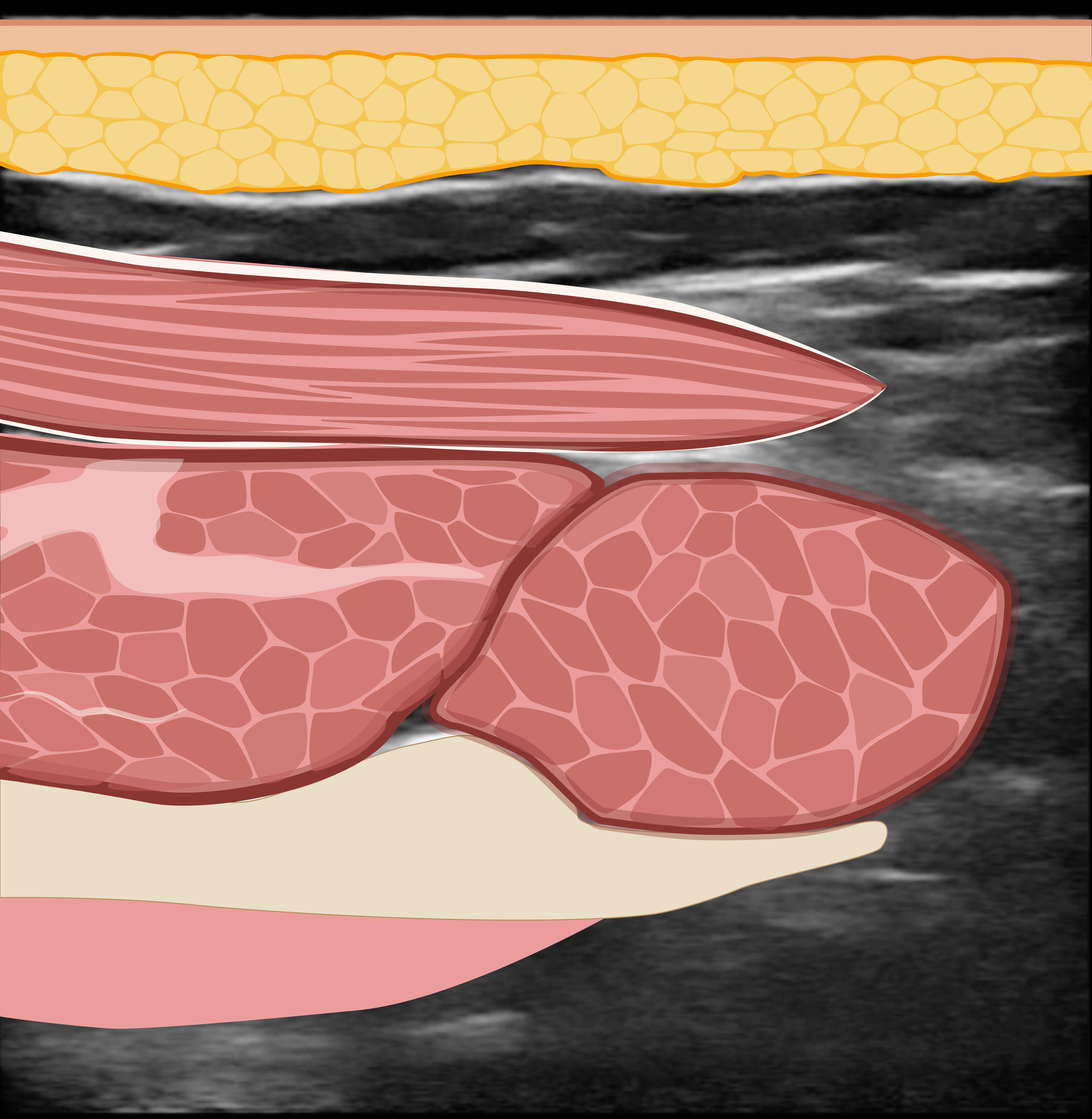
Slide the probe inferiorly in the sagittal plane across the bowl-shaped infraspinous fossa. The inferior border of the scapula can be observed as a hyperechogenic projection. The infraspinatus can be seen superior to the scapular border and the teres minor inferior to the scapular border. Both muscle bellies are in short-axis view.
REVIEW Anterior Transverse: Biceps Tendon
Use the slider to review the dynamic US technique shown through steps 1-4 to visualize the entire biceps tendon.
